
上海金畔生物科技有限公司代理AAT Bioquest荧光染料全线产品,欢迎访问AAT Bioquest荧光染料官网了解更多信息。
iFluor 568羊抗鼠免疫球蛋白(H+L)*交叉亲和 降低干扰*
 |
货号 | 16777 | 存储条件 | |
| 规格 | 1 mg | 价格 | 2544 | |
| Ex (nm) | 568 | Em (nm) | 587 | |
| 分子量 | ~150000 | 溶剂 | ||
| 产品详细介绍 | ||||
简要概述
产品基本信息
货号:16777
产品名称:iFluor 568羊抗鼠免疫球蛋白(H+L)*交叉亲和 降低干扰*
规格:1mg
储存条件:-15℃避光防潮
保质期:12个月
产品物理化学光谱特性
分子量:~150000
溶剂:水
激发波长(nm):568
发射波长(nm):587
产品介绍
iFluor 568是一种鲜红色荧光染料。 iFluor 568标记的抗IgG共轭物拥有明亮的荧光信号和良好的光稳定性。 iFluor 568共轭物的荧光强度可用于成像和流式细胞术中的稳定信号生成,在pH值为4至pH 11时对pH值不敏感。iFluor 568标记的抗体共轭物可以用K离子激光(〜568 nm)很好地激发,iFluor 568系列的光谱特性与AlexaFluor®568的光谱特性基本相同,在我们测试的相同条件下,iFluor 568抗体共轭物比相应的AlexaFluo®568更加明亮,更光稳定。这些光谱和标记特性使 iFluor 568染料家族是AlexaFluor®568的优异替代品。此外,iFluor 568二抗共轭物比相应的AlexaFluor®568标记的共轭物具有更高的信号/背景比。金畔生物是AAT Bioquest的中国代理商,为您提供最优质的iFluor 568羊抗鼠免疫球蛋白(H+L)。
点击查看光谱
图示
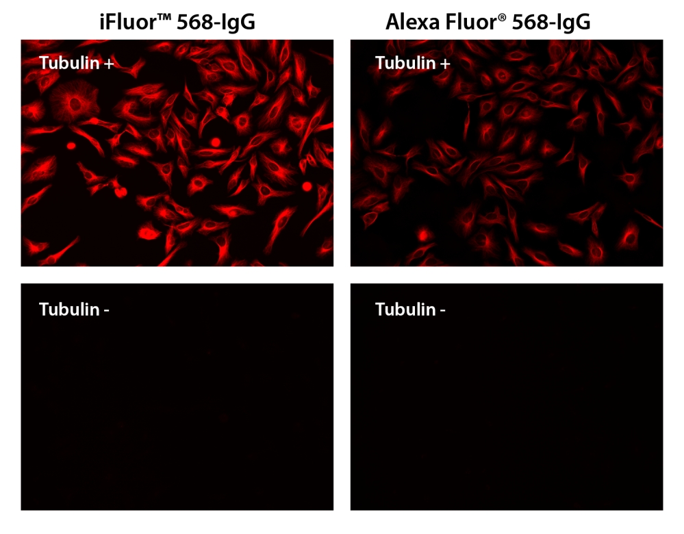
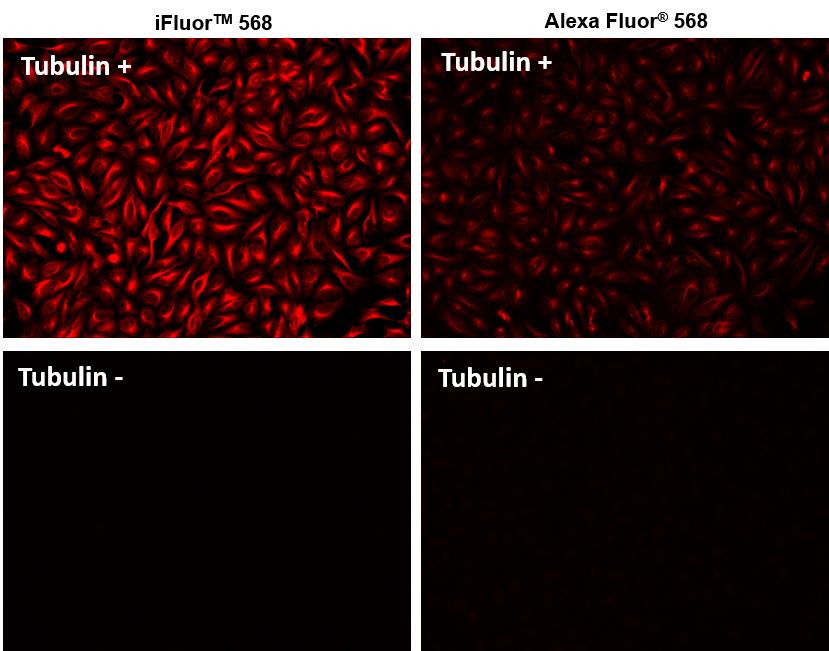
图1.将HeLa细胞与(Tubulin +)和(Tubulin-)小鼠抗微管蛋白一起孵育,然后与iFluor 568山羊抗小鼠IgG缀合物(红色,左)或AlexaFluor®568山羊抗小鼠IgG缀合物(红色 ,右)。
参考文献
Overexpression of CXCR2 predicts poor prognosis in patients with colorectal cancer.
Authors: Zhao, Jingkun and Ou, Baochi and Feng, Hao and Wang, Puxiongzhi and Yin, Shuai and Zhu, Congcong and Wang, Shenjie and Chen, Chun and Zheng, Minhua and Zong, Yaping and others
Journal: Oncotarget (2017)
Cadherin-12 enhances proliferation in colorectal cancer cells and increases progression by promoting EMT
Authors: Ma, Junjun and Zhao, Jingkun and Lu, Jun and Wang, Puxiongzhi and Feng, Hao and Zong, Yaping and Ou, Baochi and Zheng, Minhua and Lu, Aiguo
Journal: Tumor Biology (2016): 1–12
The migration and differentiation of hUC-MSCsCXCR4/GFP encapsulated in BDNF/chitosan scaffolds for brain tissue engineering
Authors: Huang, Chuanjun and Zhao, Longxiang and Gu, Jun and Nie, Dekang and Chen, Yinan and Zuo, Hao and Huan, Wei and Shi, Jinlong and Chen, Jian and Shi, Wei
Journal: Biomedical Materials (2016): 035004
Transplantation of RADA16-BDNF peptide scaffold with human umbilical cord mesenchymal stem cells forced with CXCR4 and activated astrocytes for repair of traumatic brain injury
Authors: Shi, W and Huang, CJ and Xu, XD and Jin, GH and Huang, RQ and Huang, JF and Chen, YN and Ju, SQ and Wang, Y and Shi, YW and others
Journal: Acta Biomaterialia (2016): 247–261
Antiprothrombin antibodies in a patient with secondary antiphospholipid syndrome and bleeding
Authors: Gonzalez Leon R, Garcia Hern and ez FJ, Castillo Palma MJ, Sanchez Roman J.
Journal: Med Clin (Barc) (2011): 668
Assessment of EGFR/HER2 dimerization by FRET-FLIM utilizing Alexa-conjugated secondary antibodies in relation to targeted therapies in cancers
Authors: Waterhouse BR, Gijsen M, Barber PR, Tullis ID, Vojnovic B, Kong A.
Journal: Oncotarget (2011): 728
Analysis of the effectiveness of reused primary and secondary antibodies in Western blotting analysis
Authors: Boonrod K, Roth B, Leong Ngar S, Krczal G.
Journal: Anal Biochem (2010): 124
Falsely elevated tacrolimus levels caused by immunoassay interference secondary to beta-galactosidase antibodies in an infected liver transplant recipient
Authors: Knorr JP, Grewal KS, Balasubramanian M, Young N, Zaki R, Khanmoradi K, Araya V, Ortiz J.
Journal: Pharmacotherapy (2010): 954
Nanosilver-doped DNA polyion complex membrane for electrochemical immunoassay of carcinoembryonic antigen using nanogold-labeled secondary antibodies
Authors: Wu W, Yi P, He P, Jing T, Liao K, Yang K, Wang H.
Journal: Anal Chim Acta (2010): 126
Prevalence of anti-Epstein-Barr virus antibodies in children and adolescents with secondary immunodeficiency
Authors: Buckova A.
Journal: Epidemiol Mikrobiol Imunol (2010): 133