
上海金畔生物科技有限公司代理AAT Bioquest荧光染料全线产品,欢迎访问AAT Bioquest荧光染料官网了解更多信息。
Cell Meter BX590固定化细胞活性染料
 |
货号 | 22514 | 存储条件 | 在零下15度以下保存, 避免光照 |
| 规格 | 200 Tests | 价格 | 2412 | |
| Ex (nm) | 492 | Em (nm) | 579 | |
| 分子量 | 溶剂 | DMSO | ||
| 产品详细介绍 | ||||
简要概述
产品基本信息
货号:22514
产品名称:Cell Meter BX590固定化细胞活性染料
规格:200 Tests
储存条件:保存在冰箱-15℃干燥
保质期:12个月
产品物理化学光谱特性
分子量:N/A
激发波长(nm):492
发射波长(nm):579
适用仪器
| 流式细胞仪 | |
| 激发: | 480nm激光 |
| 发射: | 575/26nm滤波片 |
| 通道: | PE通道 |
| 荧光显微镜 | |
| 激发: | Cy3/TRITC滤波片组 |
| 发射: | Cy3/TRITC滤波片组 |
| 推荐孔板: | 黑色透明 |
产品介绍
从活细胞中区分和排除死细胞可以更清晰地分离和鉴定细胞群。Cell Meter BX590固定化细胞活性染料是一类细胞不可渗透的荧光活性染料,它们通过优化以匹配常见流式细胞仪的主要激发激光,例如350、405、488、633和647 nm。这些染料不渗入活细胞,但渗入膜受损的细胞。它们与含胺和硫醇的蛋白质以及其他细胞成分发生不可逆的反应。由于具有受损膜的死细胞或固定细胞更容易与Cell Meter BX590固定化细胞活性染料发生反应,因此比具有完整膜的活细胞的染色更亮,因此这些染料可用于评估哺乳动物细胞的活与死状态。使用这些染料时,需要考虑一些因素,例如,每种染料的滴定,以确保活细胞几乎没有染色。Cell Meter BX590固定化细胞活性染料通过优化,可在488 nm的蓝色激光激发并在590 nm发射。与其他商业上类似的活性染料相比,这种Cell Meter BX590固定化细胞活性染料更加耐用、稳定。金畔生物是AAT Bioquest的中国代理商,为您提供最优质的Cell Meter BX590固定化细胞活性染料。
点击查看光谱
图示
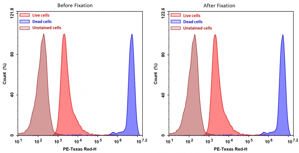
 图1.通过Cell Meter BX590固定化细胞活性染料检测Jurkat细胞活性。处理Jurkat细胞并用Cell Meter BX590(Cat#22514)染色,然后固定在3.7%甲醛中,并通过流式细胞仪进行分析。带有PE-TexasRed通道的死细胞数量(蓝峰)很容易与活细胞数量(红峰)区分开,在固定之前和之后获得的结果几乎相同。 |
参考文献
CFSE dilution to study human T and NK cell proliferation in vitro.
Authors: Terrén, Iñigo and Orrantia, Ane and Vitallé, Joana and Zenarruzabeitia, Olatz and Borrego, Francisco
Journal: Methods in enzymology (2020): 239-255
Using Carboxy Fluorescein Succinimidyl Ester (CFSE) to Identify Quiescent Glioblastoma Stem-Like Cells.
Authors: Azari, Hassan and Deleyrolle, Loic P and Reynolds, Brent A
Journal: Methods in molecular biology (Clifton, N.J.) (2018): 59-67
Estimates and impact of lymphocyte division parameters from CFSE data using mathematical modelling.
Authors: Mazzocco, Pauline and Bernard, Samuel and Pujo-Menjouet, Laurent
Journal: PloS one (2017): e0179768
Analysis of CFSE time-series data using division-, age- and label-structured population models.
Authors: Hross, Sabrina and Hasenauer, Jan
Journal: Bioinformatics (Oxford, England) (2016): 2321-9
Assessment of lymphocyte proliferation for diagnostic purpose: Comparison of CFSE staining, Ki-67 expression and 3H-thymidine incorporation.
Authors: Lašťovička, Jan and Rataj, Michal and Bartůňková, Jiřina
Journal: Human immunology (2016): 1215-1222
Poor association of allergen-specific antibody, T- and B-cell responses revealed with recombinant allergens and a CFSE dilution-based assay.
Authors: Eckl-Dorna, J and Campana, R and Valenta, R and Niederberger, V
Journal: Allergy (2015): 1222-9
Quality control of extracorporeal photochemotherapy: Proliferation assay using CFSE validated according to ISO 15189:2007 standards.
Authors: Faivre, Lionel and Lecouflet, Lucie and Liu, Wang-Qing and Khadher, Isabelle and Lahaie, Camille and Vidal, Michel and Legouvello, Sabine and Beaumont, Jean-Louis and Bierling, Philippe and Rouard, Hélène and Birebent, Brigitte
Journal: Cytometry. Part B, Clinical cytometry (2015): 30-9
Mathematical models for CFSE labelled lymphocyte dynamics: asymmetry and time-lag in division.
Authors: Luzyanina, Tatyana and Cupovic, Jovana and Ludewig, Burkhard and Bocharov, Gennady
Journal: Journal of mathematical biology (2014): 1547-83
Quality control of extracorporeal photochemotherapy: Proliferation assay using CFSE validated according to ISO 15189:2007 standards.
Authors: Lionel, Faivre and Lucie, Lecouflet and Wang-Qing, Liu and Isabelle, Khadher and Camille, Lahaie and Michel, Vidal and Sabine, Legouvello and Jean-Louis, Beaumont and Philippe, Bierling and Hélène, Rouard and Brigitte, Birebent
Journal: Cytometry. Part B, Clinical cytometry (2014)
Asymmetry of Cell Division in CFSE-Based Lymphocyte Proliferation Analysis.
Authors: Bocharov, Gennady and Luzyanina, Tatyana and Cupovic, Jovana and Ludewig, Burkhard
Journal: Frontiers in immunology (2013): 264