上海金畔生物科技有限公司代理AAT Bioquest荧光染料全线产品,欢迎访问AAT Bioquest荧光染料官网了解更多信息。
Cell Meter 细胞自噬检测荧光成像试剂盒
 |
货号 | 23001 | 存储条件 | 在零下15度以下保存, 避免光照 |
| 规格 | 200 Tests | 价格 | 3840 | |
| Ex (nm) | 333 | Em (nm) | 518 | |
| 分子量 | 溶剂 | |||
| 产品详细介绍 | ||||
简要概述
自噬是动物细胞内大分子降解的主要途径之一。 自噬过程涉及将细胞质和细胞内细胞器隔离在称为自噬体的膜结合液泡中,将自噬体与溶酶体融合,然后降解被隔离的物质。 Cell Meter 细胞自噬检测荧光成像试剂盒采用Autophagy Super Blue 作为特异性自噬标记物来分析自噬活性。 该测定法经过优化,可直接检测分离和粘附细胞中的自噬。 Cell Meter 细胞自噬检测荧光成像试剂盒针对荧光显微镜进行了优化。 它提供了比其他市售自噬探针更高的选择性。金畔生物是AAT Bioquest的中国代理商,为您提供最优质的Cell Meter 细胞自噬检测荧光成像试剂盒。
适用仪器
| 荧光显微镜 | |
| 激发: | DAPI滤波片 |
| 发射: | DAPI滤波片 |
| 推荐孔板: | 黑色透明 |
产品说明书
样品实验方案
简要概述
1.用您的测试化合物以1-2×10⁴个细胞/孔的密度制备细胞
2.添加自噬Super Blue 工作溶液
3.在37°C孵育15-60分钟
4.用洗涤缓冲液洗涤细胞
5.使用DAPI滤光片组检测在Ex / Em = 330/520 nm(截止= 475 nm)处的荧光
溶液配制
工作溶液配制
将20μL500X自噬Super Blue (组分A)添加到10 mL染色缓冲液(组分B)中,并充分混合以制成Autophagy Super Blue 工作溶液,避光。 注意:20μL500X自噬Super Blue (组分A)足以容纳一个96孔板。
实验步骤
1.根据您的特定诱导方案将细胞培养至最适合自噬诱导的密度(约1-2×10⁴细胞/孔/ 96孔板)。同时,在每种标记条件下,以与诱导种群相同的密度培养非诱导的阴性对照细胞种群。
2.除去培养基。
3.在每孔中加入100 µL /孔(96孔板)或25 µL /孔(384孔板)的自噬Super Blue 工作溶液。
4.将细胞在37°C,5%CO2培养箱中孵育15至60分钟。注意:适当的孵育时间取决于所用的单个细胞类型和细胞浓度。
5.用洗涤缓冲液(组分C)洗涤细胞3-4次,然后向每个孔中添加100 µL洗涤缓冲液(组分C)。注意:建议增加标记浓度或孵育时间,使染料在细胞未充分染色的情况下积累。
6.用荧光酶标仪在Ex / Em = 330/520 nm(Cutoff = 475 nm)上检测荧光强度,或使用带有DAPI滤光片组的荧光显微镜。
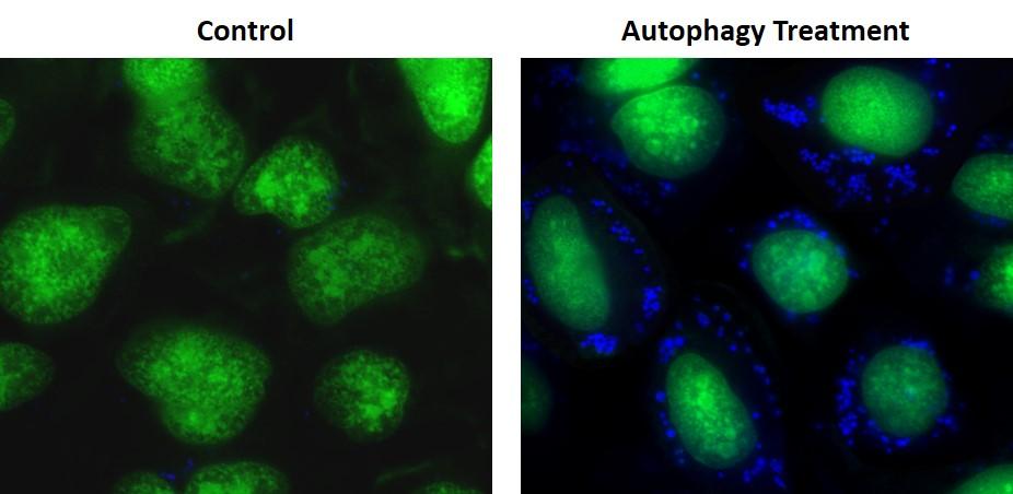
图示
 图1.通过自噬在HeLa细胞中诱导自噬Super Blue 标记的囊泡。 将HeLa细胞在常规DMEM培养基(左:对照)中或在含5%血清的1X HBSS缓冲液中(右:自噬处理)孵育16小时。对照细胞和处理过的细胞均在37°C,5%CO2培养箱中与Autophagy Super Blue 工作溶液一起孵育20分钟,并用洗涤缓冲液洗涤3次。 立即在带有DAPI通道(蓝色)的荧光显微镜下对细胞成像。 细胞核用Nuclear Gree LCS1(绿色)染色。 |
参考文献
Methods for Measuring Autophagy Levels in Disease
Authors: Phadwal, Kanchan and Kurian, Dominic
Journal: (2017): 195–211
High glucose induces bone marrow-derived mesenchymal stem cell senescence by upregulating autophagy
Authors: Chang, Tzu-Ching and Hsu, Min-Fen and Wu, Kenneth K
Journal: PloS one (2015): e0126537
Licochalcone A induces autophagy through PI3K/Akt/mTOR inactivation and autophagy suppression enhances Licochalcone A-induced apoptosis of human cervical cancer cells
Authors: Tsai, Jen-Pi and Lee, Chien-Hsing and Ying, Tsung-Ho and Lin, Chu-Liang and Lin, Chia-Liang and Hsueh, Jung-Tsung and Hsieh, Yi-Hsien
Journal: Oncotarget (2015): 28851
An autophagy inhibitor enhances the inhibition of cell proliferation
Authors: Yao F, Wang G, Wei W, Tu Y, Tong H, Sun S.
Journal: Mol Med Report (2012): 84
High-Throughput Screening for AntiInfluenza A Virus Drugs and Study of the Mechanism of Procyanidin on Influenza A VirusInduced Autophagy
Authors: Dai J, Wang G, Li W, Zhang L, Yang J, Zhao X, Chen X, Xu Y, Li K.
Journal: J Biomol Screen. (2012)
Tgf-beta1 induces autophagy and promotes apoptosis in renal tubular epithelial cells
Authors: Xu Y, Yang S, Huang J, Ruan S, Zheng Z, Lin J.
Journal: Int J Mol Med. (2012)
beta-Elemene induces apoptosis as well as protective autophagy in human non-small-cell lung cancer A549 cells
Authors: Liu J, Hu XJ, Jin B, Qu XJ, Hou KZ, Liu YP.
Journal: J Pharm Pharmacol (2012): 146
A new fluorescence-based assay for autophagy
Authors: Proikas-Cezanne T, Codogno P.
Journal: Chem Biol (2011): 940
Inhibition of induced autophagy increases apoptosis of Nara-H cells
Authors: Nakamura O, Hitora T, Akisue T, Kawamoto T, Yamagami Y, Yamamoto T.
Journal: Int J Oncol (2011): 1545
Reactive oxygen species contribute to oridonin-induced apoptosis and autophagy in human cervical carcinoma HeLa cells
Authors: Zhang YH, Wu YL, Tashiro S, Onodera S, Ikejima T.
Journal: Acta Pharmacol Sin (2011): 1266