
上海金畔生物科技有限公司代理AAT Bioquest荧光染料全线产品,欢迎访问AAT Bioquest荧光染料官网了解更多信息。
Cell Meter 磷脂酰丝氨酸凋亡检测试剂盒 绿色荧光,适合微孔板检测
 |
货号 | 22791 | 存储条件 | 在2-8度冷藏保存, 避免光照 |
| 规格 | 100 Tests | 价格 | 2544 | |
| Ex (nm) | 498 | Em (nm) | 520 | |
| 分子量 | 溶剂 | |||
| 产品详细介绍 | ||||
简要概述
Cell Meter检测试剂盒是一类用于检测细胞功能的系列工具,包括细胞活性、细胞毒性、细胞凋亡、细胞膜电位以及细胞周期等方面的指标。每种检测方案均能提供不同荧光颜色的检测方案。这些高效的检测方案为从多角度研究细胞功能活动提供了一种十分有效的方法。Cell Meter磷脂酰丝氨酸凋亡检测试剂盒 *绿色荧光,适合微孔板检测*是通过检测PS的翻转来测定细胞凋亡。在凋亡细胞中,PS从胞内移位到胞外表面,因此PS出现在细胞的外表面可以作为细胞凋亡的起始或者中间阶段的通用指示器,也可以通过其形态学改变来测定。该试剂盒使用我们独有的绿色荧光Apopxin PS传感器特异性的结合PS,其亲和性高于Annexin V(Kd<10nM)。该试剂盒使用的PS传感器结合膜上的PS后,发出绿色的荧光。因为与PS的高亲和性,该试剂盒比其它基于Annexin-V,仅能用荧光显微镜和流式检测凋亡的试剂盒更稳定,并且本试剂盒除上述两种仪器外还能使用荧光酶标仪。
点击查看细胞样品制备
适用仪器
| 荧光酶标仪 | |
| 激发: | 490nm |
| 发射: | 525nm |
| cutoff: | 515nm |
| 推荐孔板: | 黑色透明 |
| 读取模式: | 底读模式 |
产品说明书
样品实验方案
简要概述
1.用测试化合物制备细胞(100 µL /孔/ 96孔板或25 µL /孔/ 384孔板)
2.加入等量的Apopxin™Green工作溶液
3.在室温下孵育1小时
4.在Ex / Em = 490/525 nm(截止= 515 nm)或带有FITC滤光片的荧光显微镜下监控荧光强度(底部读取模式)
溶液配制
工作溶液配制
将10 µL 100X Apopxin™Green(组分A)添加到1 mL的测定缓冲液(组分B)中,并充分混合以制成Apopxin™Green工作溶液。
操作步骤
1.通过向细胞制备缓冲液中加入10XL /孔(96孔板)的10X测试化合物溶液,以所需的测试化合物处理细胞,总体积为100μL/孔。对于384孔板,请在细胞制备缓冲液中使用5 µL /孔的5X测试化合物溶液,总体积为25 µL /孔。对于空白孔(不含细胞的培养基),添加相同量的化合物缓冲液。
2.将细胞板在5%CO2、37°C的培养箱中孵育所需的时间(对于喜树碱处理过的Jurkat细胞,需要4-6小时)以诱导凋亡。
3.在每个孔中添加100 µL /孔(96孔板)或25 µL /孔(384孔板)的Apopxin™Green工作溶液。
4.在避光条件下,于室温下孵育细胞板至少1小时。
5.以800 rpm的速度离心细胞板(特别是非粘附细胞)2分钟(制动)。
6.使用荧光酶标仪(Ext / Em = 490/525 nm(Cutoff = 515 nm))或使用带有FITC滤光片的荧光显微镜的图像池监控荧光强度。
图示

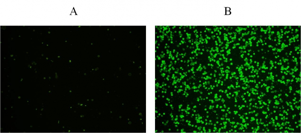
图1.在Costar黑色壁/透明底部96孔板中,用Cell Meter™磷脂酰丝氨酸凋亡检测试剂盒染色的Jurkat细胞图像。 A:未经处理的对照细胞。 B:用20 µM喜树碱处理5小时的细胞。
参考文献
Physiological effects of the herbicide glyphosate on the cyanobacterium Microcystis aeruginosa
Authors: Wu, Liang and Qiu, Zhihao and Zhou, Ya and Du, Yuping and Liu, Chaonan and Ye, Jing and Hu, Xiaojun
Journal: Aquatic Toxicology (2016): 72–79
Tumor-selective mitochondrial network collapse induced by atmospheric gas plasma-activated medium.
Authors: Saito, Kosuke and Asai, Tomohiko and Fujiwara, Kyoko and Sahara, Junki and Koguchi, Haruhisa and Fukuda, Noboru and Suzuki-Karasaki, Miki and Soma, Masayoshi and Suzuki-Karasaki, Yoshihiro
Journal: Oncotarget (2016)
Inhibition of malignant phenotypes of human osteosarcoma cells by a gene silencer, a pyrrole–imidazole polyamide, which targets an E-box motif
Authors: Taniguchi, Masashi and Fujiwara, Kyoko and Nakai, Yuji and Ozaki, Toshinori and Koshikawa, Nobuko and Toshio, Kojima and Kataba, Motoaki and Oguni, Asako and Matsuda, Hiroyuki and Yoshida, Yukihiro and others
Journal: FEBS open bio (2014): 328–334
Detection of apoptosis based on the interaction between annexin V and phosphatidylserine
Authors: Liu T, Zhu W, Yang X, Chen L, Yang R, Hua Z, Li G.
Journal: Anal Chem (2009): 2410
Evaluation of cell surface expression of phosphatidylserine in ovarian carcinoma effusions using the annexin-V/7-AAD assay: clinical relevance and comparison with other apoptosis parameters
Authors: Dong HP, Holth A, Kleinberg L, Ruud MG, Elstr and MB, Trope CG, Davidson B, Risberg B.
Journal: Am J Clin Pathol (2009): 756
Mobilization of lysosomal calcium regulates the externalization of phosphatidylserine during apoptosis
Authors: Mirnikjoo B, Balasubramanian K, Schroit AJ.
Journal: J Biol Chem (2009): 6918
Peptidic targeting of phosphatidylserine for the MRI detection of apoptosis in atherosclerotic plaques
Authors: Burtea C, Laurent S, Lancelot E, Ballet S, Murariu O, Rousseaux O, Port M, V and er Elst L, Corot C, Muller RN.
Journal: Mol Pharm (2009): 1903
Suicidal membrane repair regulates phosphatidylserine externalization during apoptosis
Authors: Mirnikjoo B, Balasubramanian K, Schroit AJ.
Journal: J Biol Chem (2009): 22512
Trivalent methylated arsenical-induced phosphatidylserine exposure and apoptosis in platelets may lead to increased thrombus formation
Authors: Bae ON, Lim KM, Noh JY, Chung SM, Kim SH, Chung JH.
Journal: Toxicol Appl Pharmacol (2009): 144
Discovery of a phosphatidylserine-recognizing peptide and its utility in molecular imaging of tumour apoptosis
Authors: Thapa N, Kim S, So IS, Lee BH, Kwon IC, Choi K, Kim IS.
Journal: J Cell Mol Med (2008): 1649