Description: The BM8 monoclonal antibody reacts with mouse F4/80 antigen, an approximay 125 kDa transmembrane protein. The F4/80 antigen is expressed by a majority of mature macrophages and is the best marker for this population of cells. However, other cell types such as Langerhans cells and liver Kupffer cells have been reported to express this antigen. Expression of F4/80 commences during early myeloid development and is upregulated on all BM cells stimulated in vitro with M-CSF. It has been shown that some cytokines downregulate the expression of F4/80 resulting in lack of F4/80 antigen on a subpopulation of macrophages, especially in the lymphoid microenvironment in vivo
DESCRIPTION Source E. coliderived Thr22Ala158 Accession # Q62386.1 Nterminal Sequence Analysis Thr22 Structure / Form Disulfidelinked homodimer Predicted Molecular Mass 15.5 kDa (monomer) SPECIFICATIONS Activity Measured by its ability to induce IL6 secretion by NIH-3T3 mouse embryonic fibroblast cells. Yao, Z. et al. (1995) Immunity 3:811. The ED50 for this effect is typically 0.251.25 ng/mL. Endotoxin Level <1.0 EU per 1 μg of the protein by the LAL method. Purity >97%, by SDSPAGE under reducing conditions and visualized by silver stain. Formulation Lyophilized from a 0.2 μm filtered solution in Acetonitrile and TFA with BSA as a carrier protein. See Certificate of Analysis for details. PREPARATION AND STORAGE Reconstitution Reconstitute at 25 μg/mL in sterile 4 mM HCl containing at least 0.1% human or bovine serum albumin. Shipping The product is shipped at ambient temperature. Upon receipt, store it immediay at the temperature recommended below. Stability & Storage Use a manual defrost freezer and avoid repeated freezethaw cycles. l 12 months from date of receipt, 20 to 70 °C as supplied. l 1 month, 2 to 8 °C under sterile conditions after reconstitution. l 3 months, 20 to 70 °C under sterile conditions after reconstitution. BACKGROUND Interleukin 17 (also known as CTLA8) is a T cellexpressed pleiotropic cytokine that exhibits a high degree of homology to a protein encoded by the ORF13 gene of herpesvirus Saimiri. cDNA clones encoding IL17 have been isolated from activated rat, mouse and human T cells. Mouse IL17 cDNA encodes a 158 amino acid (aa) residue precursor protein with a 21 amino acid residue signal peptide that is cleaved to yield the 137 aa residue mature IL17. Both recombinant and natural IL17 have been shown to exist as disulfide linked homodimers. At the amino acid level, mIL17 shows 57% and 87% sequence identity with herpesvirus and rat IL17, respectively. An IL17 specific mouse cell surface receptor (IL17 R) has been cloned. While the expression of IL17 mRNA is restricted to activated alpha beta TCR+CD4CD8T cells, the expression of mIL17 R mRNA has been detected in virtually all cells and tissues tested. IL17 exhibits multiple biological activities on a variety of cells including: the induction of IL6 and IL8 production in fibroblasts? the enhancement of surface expression of ICAM1 in fibroblasts? activation of NFκB and costimulation of T cell proliferation. References: 1. Kennedy, J. et al., (1996) J. Interferon Cytokine Res. 16:611. 2. Yao, Z. et al., (1995) J. Immunol. 155:5483. 3. Yao, Z. et al., (1995) Immunity 3:811. 4. Rouvier, E. et al., (1993) J. Immunol. 150:5445.
Signal Transduction >> Cytoskeleton / ECM >> Cytoskeleton >> Intermediate Filaments >> Class I >> Cytokeratins
Datasheet PDF
SDS
Immunohistochemistry (Formalin/PFA-fixed paraffin-embedded sections) – pan Cytokeratin antibody [PCK-26] (ab6401)(enlarge)
Immunohistochemistry (Formalin/PFA-fixed paraffin-embedded sections) – pan Cytokeratin antibody [PCK-26] (ab6401)(enlarge)
Western blot – pan Cytokeratin antibody [PCK-26] (ab6401)(enlarge)
Applications
Show applications key
Our Abpromise guarantee covers the use of ab6401 in the following tested applications.
The application notes include recommended starting dilutions; optimal dilutions/concentrations should be determined by the end user.
ShowHide4 Images
WB
Four stars(5 Abreviews)WB: Use at an assay depe…Read more →
WB: Use at an assay dependent dilution.
ShowHide
IHC-FoFr
IHC-FoFr: Use at an assa…Read more →
IHC-FoFr: Use at an assay dependent dilution.
ShowHide3 Images
IHC-P
Five stars(3 Abreviews)IHC-P: Use at an assay d…Read more →
IHC-P: Use at an assay dependent dilution.
ShowHide2 Images
IHC-Fr
Four stars(3 Abreviews)IHC-Fr: Use at an assay …Read more →
IHC-Fr: Use at an assay dependent dilution.
IF
IF: 1/300.
IF: 1/300.
ShowHide
Dot Blot
Dot: Use at an assay dep…Read more →
Dot: Use at an assay dependent dilution.
ShowHide
Flow Cyt
See more…Read more →
Flow Cyt: Use at an assay dependent dilution. PubMed: 19855980Use 10µl for 5 x 10⁷ cells.
2 Images
ICC/IF
Five stars(2 Abreviews)ICC/IF: 1/300.
ICC/IF: 1/300.
Target
Relevance
Cytokeratins, a group comprising at least 29 different proteins, are characteristic of epithelial and trichocytic cells. Cytokeratins 1, 4, 5, 6, and 8 are members of the type II neutral to basic subfamily. Monoclonal anti cytokeratins are specific markers of epithelial cell differentiation and have been widely used as tools in tumor identification and classification. Monoclonal Anti Pan Cytokeratin (mixture) is a broadly reactive reagent, which recognizes epitopes present in most human epithelial tissues. It facilitates typing of normal, metaplastic and neoplastic cells. Synergy between the various components results in staining amplification. This enables identification of cells, which would otherwise be stained only marginally. The mixture may aid in the discrimination of carcinomas and nonepithelial tumors such as sarcomas, lymphomas and neural tumors. It is also useful in detecting micrometastases in lymph nodes, bone marrow and other tissues and for determining the origin of poorly differentiated tumors. There are two types of cytokeratins the acidic type I cytokeratins and the basic or neutral type II cytokeratins. Cytokeratins are usually found in pairs comprising a type I cytokeratin and a type II cytokeratin. Usually the type II cytokeratins are 8kD larger than their type I counterparts.
Cellular localization
Cytoplasmic
Anti-pan Cytokeratin antibody [PCK-26] images:
Immunohistochemistry (Formalin/PFA-fixed paraffin-embedded sections) – pan Cytokeratin antibody [PCK-26] (ab6401)
ab6401 staining pan Cytokeratin in Mouse liver tumor tissue sections by IHC-P (Paraformaldehyde fixed paraffin embedded sections). Tissue was fixed with paraformaldehyde and blocked with maleate buffer blocking solution for 30 minutes and 22°C. Antigen retrieval was by heat mediation in citrate buffer. Samples were incubated with primary antibody (1/500) in maleate buffer blocking solution for 16 hours at 22°C. An undiluted biotin-conjugated Donkey polyclonal to mouse IgG was used as secondary antibody.
This image is courtesy of an Abreview submitted by Dr Asha Seth
See Abreview
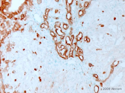
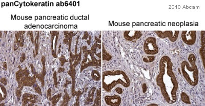
Immunohistochemistry (Formalin/PFA-fixed paraffin-embedded sections) – pan Cytokeratin antibody [PCK-26] (ab6401)
ab6401 staining pan Cytokeratin in mouse pancreatic ductal adenocarcinoma (left-hand panel) and mouse pancreatic neoplasia (right-hand panel) sections by immunohistochemistry (IHC-P – paraformaldehyde-fixed, paraffin-embedded sections). Tissue samples were fixed with paraformaldehyde and blocked with 10% serum for 1 hour at room temperature; heat mediated antigen retrieval was performed. The sample was incubated with primary antibody (1/250) at 4°C for 8 hours. A Biotin-conjugated Goat polyclonal (1/1000) was used as the secondary antibody.
This image is courtesy of an anonymous Abreview
See Abreview
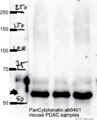
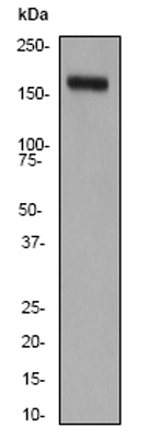
Western blot – pan Cytokeratin antibody [PCK-26] (ab6401)
Observed band size : 55 kDa (why is the actual band size different from the predicted?)
All lanes: pan Cytokeratin antibody (ab6401) at 1/1000 dilution + whole cell lysate of Primary mouse pancreatic cancer cell lines (35 µg)
Secondary: An HRP-conjugated Sheep anti-mouse IgG polyclonal (1/5000) developed using the ECL technique
Performed under non-reducing conditions.
Blocking Step: 10% Milk for 1 hour at room temperature
This image is courtesy of an Abreview submitted by Pawel Mazur
See Abreview
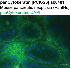
Immunohistochemistry (Frozen sections) – pan Cytokeratin antibody [PCK-26] (ab6401)
ab6401 staining pan Cytokeratin in Mouse pancreatic neoplasia tissue sections by Immunohistochemistry (IHC-Fr – frozen sections). Tissue was fixed with paraformaldehyde and blocked with 1% BSA for 1 hour at room temperature. Samples were incubated with primary antibody (1/250 in PBS) for 8 hours at 4°C. An Alexa Fluor®488-conjugated goat anti-mouse IgG polyclonal (1/1000) was used as the secondary antibody.
This image is courtesy of an anonymous Abreview
See Abreview
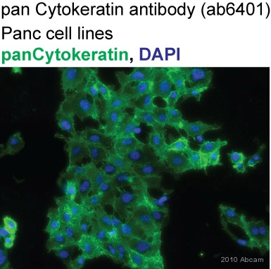
Immunocytochemistry/ Immunofluorescence – pan Cytokeratin antibody [PCK-26] (ab6401)
ab6401 staining pan Cytokeratin in Human Panc1 cells by ICC/IF (Immunocytochemistry/immunofluorescence). Cells were fixed with acetone/methanol (1:1) and blocked with 1% BSA for 1 hour at room temperature. Samples were incubated with primary antibody (1/300 in PBS) for 2 hours. An Alexa Fluor®488-conjugated DGoat anti-mouse IgG polyclonal (1/1000) was used as the secondary antibody. Nuclei were counterstained with DAPI.
This image is courtesy of an anonymous Abreview
See Abreview
References for Anti-pan Cytokeratin antibody [PCK-26] (ab6401)
MCSF, also known as CSF1, is a fourαhelicalbundle cytokine that is the primary regulator of macrophage survival, proliferation and differentiation (1 3). MCSF is also essential for the survival and proliferation of osteoclast progenitors (1, 4). MCSF also primes and enhances macrophage killing of tumor cells and microorganisms, regulates the release of cytokines and other inflammatory modulators from macrophages, and stimulates pinocytosis (2, 3). MCSF increases during pregnancy to support implantation and growth of the decidua and placenta (5). Sources of MCSF include fibroblasts, activated macrophages, endometrial secretory epithelium, bone marrow stromal cells and activated endothelial cells (1 5). The MCSF receptor (cfms) transduces its pleotropic effects and mediates its endocytosis. MCSF mRNAs of various sizes occur (3 9). Full length mouse MCSF transcripts encode a 520 amino acid (aa) type I transmembrane (TM) protein with a 462 aa extracellular region, a 21 aa TM domain, and a 37 aa cytoplasmic tail that forms a 140 kDa covalent dimer. Differential processing produces two proteolytically cleaved, secreted dimers. One is an Nand Oglycosylated 86 kDa dimer, while the other is modified by both glycosylation and chondroitinsulfate proteoglycan (PG) to generate a 200 kDa subunit. Although PGmodified MCSF can circulate, it may be immobilized by attachment to type V collagen (8). Shorter transcripts encode M CSF that lacks cleavage and PG sites and produces an Nglycosylated 68 kDa TM dimer and a slowly produced 44 kDa secreted dimer (7). Although forms may vary in activity and halflife, all contain the Nterminal 150 aa portion that is necessary and sufficient for interaction with the M CSF receptor (10, 11). The first 229 aa of mature mouse MCSF shares 87%, 83%, 82% and 81% aa identity with corresponding regions of rat, dog, cow and human MCSF, respectively (12, 13). Human MCSF is active in the mouse, but mouse MCSF is reported to be speciesspecific


 (enlarge)
 (enlarge)
 (enlarge)
 (enlarge)
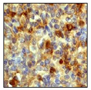 (enlarge)
 (enlarge)