详细描述: CD21 (also known as complement receptor (CR) type 2) is a type I integral membrane glycoprotein that serves as a receptor for the C3d complement fragment and for the Epstein-Barr virus. It plays a role in B cell activation and proliferation and undergoes
Delivery dates and prices will be calculated based on your ZIP
Items in stock will be shipped on 04/22/2019
I cannot find my country in this list
Free express shipping from $400!
Add products for to your cart and get free express shipping Your cart total is $0 , and your order qualifies for free express shipping to , ZIP .
Delivery options for stock items to , ZIP
Items in stock will be shipped on 04/22/2019
Change country or ZIP
Free express shipping from $400!
Add products for to your cart and get free express shipping to , ZIP .
Free express shipping from $400!
Your cart total is $0, and your order qualifies for free express shipping to , ZIP .
Change country or ZIP
Cyanine5 maleimide is a mono-reactive dye which selectively couples with thiol groups (for example, with cysteines in peptides and proteins) to give labeled conjugates.
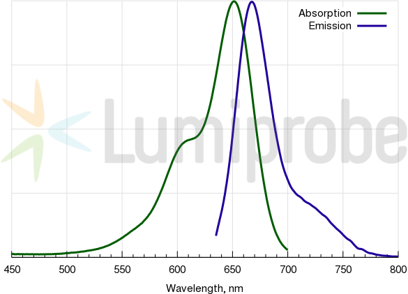
Cyanine5 is an analog of Cy5®, a common fluorophore which is compatible with various instrumentation like microscopes, imagers, and fluorescence readers.
For the labeling of antibodies and sensitive proteins we recommend to use the water soluble sulfo-Cyanine5 maleimide.
Download in ChemDraw format
Cy5 excitation and emission spectra
Recommended protocol
Maleimide labeling of proteins and other thiolated biomolecules
Customers also purchased with this product
Cyanine5 DBCO
Cyanine5 dye with dibenzocyclooctyne (DBCO) functional group for copper free Click chemistry.
Signal Transduction >> Cytoskeleton / ECM >> Cytoskeleton >> Intermediate Filaments >> Class I >> Cytokeratins
Datasheet PDF
SDS
Immunohistochemistry (Formalin/PFA-fixed paraffin-embedded sections) – pan Cytokeratin antibody [PCK-26] (ab6401)(enlarge)
Immunohistochemistry (Formalin/PFA-fixed paraffin-embedded sections) – pan Cytokeratin antibody [PCK-26] (ab6401)(enlarge)
Western blot – pan Cytokeratin antibody [PCK-26] (ab6401)(enlarge)
Applications
Show applications key
Our Abpromise guarantee covers the use of ab6401 in the following tested applications.
The application notes include recommended starting dilutions; optimal dilutions/concentrations should be determined by the end user.
ShowHide4 Images
WB
Four stars(5 Abreviews)WB: Use at an assay depe…Read more →
WB: Use at an assay dependent dilution.
ShowHide
IHC-FoFr
IHC-FoFr: Use at an assa…Read more →
IHC-FoFr: Use at an assay dependent dilution.
ShowHide3 Images
IHC-P
Five stars(3 Abreviews)IHC-P: Use at an assay d…Read more →
IHC-P: Use at an assay dependent dilution.
ShowHide2 Images
IHC-Fr
Four stars(3 Abreviews)IHC-Fr: Use at an assay …Read more →
IHC-Fr: Use at an assay dependent dilution.
IF
IF: 1/300.
IF: 1/300.
ShowHide
Dot Blot
Dot: Use at an assay dep…Read more →
Dot: Use at an assay dependent dilution.
ShowHide
Flow Cyt
See more…Read more →
Flow Cyt: Use at an assay dependent dilution. PubMed: 19855980Use 10µl for 5 x 10⁷ cells.
2 Images
ICC/IF
Five stars(2 Abreviews)ICC/IF: 1/300.
ICC/IF: 1/300.
Target
Relevance
Cytokeratins, a group comprising at least 29 different proteins, are characteristic of epithelial and trichocytic cells. Cytokeratins 1, 4, 5, 6, and 8 are members of the type II neutral to basic subfamily. Monoclonal anti cytokeratins are specific markers of epithelial cell differentiation and have been widely used as tools in tumor identification and classification. Monoclonal Anti Pan Cytokeratin (mixture) is a broadly reactive reagent, which recognizes epitopes present in most human epithelial tissues. It facilitates typing of normal, metaplastic and neoplastic cells. Synergy between the various components results in staining amplification. This enables identification of cells, which would otherwise be stained only marginally. The mixture may aid in the discrimination of carcinomas and nonepithelial tumors such as sarcomas, lymphomas and neural tumors. It is also useful in detecting micrometastases in lymph nodes, bone marrow and other tissues and for determining the origin of poorly differentiated tumors. There are two types of cytokeratins the acidic type I cytokeratins and the basic or neutral type II cytokeratins. Cytokeratins are usually found in pairs comprising a type I cytokeratin and a type II cytokeratin. Usually the type II cytokeratins are 8kD larger than their type I counterparts.
Cellular localization
Cytoplasmic
Anti-pan Cytokeratin antibody [PCK-26] images:
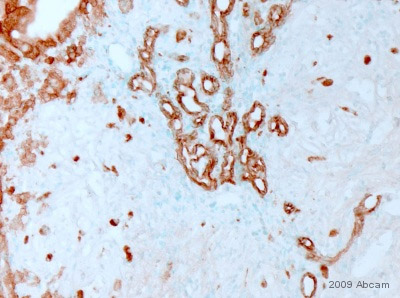
Immunohistochemistry (Formalin/PFA-fixed paraffin-embedded sections) – pan Cytokeratin antibody [PCK-26] (ab6401)
ab6401 staining pan Cytokeratin in Mouse liver tumor tissue sections by IHC-P (Paraformaldehyde fixed paraffin embedded sections). Tissue was fixed with paraformaldehyde and blocked with maleate buffer blocking solution for 30 minutes and 22°C. Antigen retrieval was by heat mediation in citrate buffer. Samples were incubated with primary antibody (1/500) in maleate buffer blocking solution for 16 hours at 22°C. An undiluted biotin-conjugated Donkey polyclonal to mouse IgG was used as secondary antibody.
This image is courtesy of an Abreview submitted by Dr Asha Seth
See Abreview
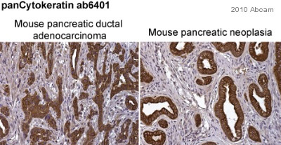
Immunohistochemistry (Formalin/PFA-fixed paraffin-embedded sections) – pan Cytokeratin antibody [PCK-26] (ab6401)
ab6401 staining pan Cytokeratin in mouse pancreatic ductal adenocarcinoma (left-hand panel) and mouse pancreatic neoplasia (right-hand panel) sections by immunohistochemistry (IHC-P – paraformaldehyde-fixed, paraffin-embedded sections). Tissue samples were fixed with paraformaldehyde and blocked with 10% serum for 1 hour at room temperature; heat mediated antigen retrieval was performed. The sample was incubated with primary antibody (1/250) at 4°C for 8 hours. A Biotin-conjugated Goat polyclonal (1/1000) was used as the secondary antibody.
This image is courtesy of an anonymous Abreview
See Abreview
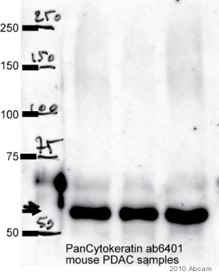
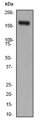
Western blot – pan Cytokeratin antibody [PCK-26] (ab6401)
Observed band size : 55 kDa (why is the actual band size different from the predicted?)
All lanes: pan Cytokeratin antibody (ab6401) at 1/1000 dilution + whole cell lysate of Primary mouse pancreatic cancer cell lines (35 µg)
Secondary: An HRP-conjugated Sheep anti-mouse IgG polyclonal (1/5000) developed using the ECL technique
Performed under non-reducing conditions.
Blocking Step: 10% Milk for 1 hour at room temperature
This image is courtesy of an Abreview submitted by Pawel Mazur
See Abreview
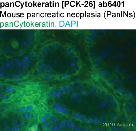
Immunohistochemistry (Frozen sections) – pan Cytokeratin antibody [PCK-26] (ab6401)
ab6401 staining pan Cytokeratin in Mouse pancreatic neoplasia tissue sections by Immunohistochemistry (IHC-Fr – frozen sections). Tissue was fixed with paraformaldehyde and blocked with 1% BSA for 1 hour at room temperature. Samples were incubated with primary antibody (1/250 in PBS) for 8 hours at 4°C. An Alexa Fluor®488-conjugated goat anti-mouse IgG polyclonal (1/1000) was used as the secondary antibody.
This image is courtesy of an anonymous Abreview
See Abreview
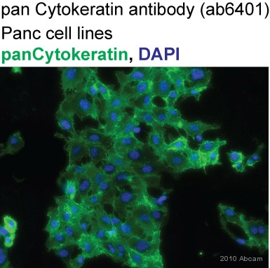
Immunocytochemistry/ Immunofluorescence – pan Cytokeratin antibody [PCK-26] (ab6401)
ab6401 staining pan Cytokeratin in Human Panc1 cells by ICC/IF (Immunocytochemistry/immunofluorescence). Cells were fixed with acetone/methanol (1:1) and blocked with 1% BSA for 1 hour at room temperature. Samples were incubated with primary antibody (1/300 in PBS) for 2 hours. An Alexa Fluor®488-conjugated DGoat anti-mouse IgG polyclonal (1/1000) was used as the secondary antibody. Nuclei were counterstained with DAPI.
This image is courtesy of an anonymous Abreview
See Abreview
References for Anti-pan Cytokeratin antibody [PCK-26] (ab6401)


 (enlarge)
 (enlarge)
 (enlarge)
 (enlarge)
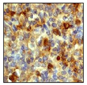 (enlarge)
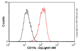 (enlarge)