
上海金畔生物科技有限公司代理AAT Bioquest荧光染料全线产品,欢迎访问AAT Bioquest荧光染料官网了解更多信息。
Cell Meter 线粒体膜电位检测试剂盒 橙色荧光 适合微孔板检测
 |
货号 | 22805 | 存储条件 | 在零下15度以下保存, 避免光照 |
| 规格 | 500 Tests | 价格 | 2544 | |
| Ex (nm) | 546 | Em (nm) | 575 | |
| 分子量 | 溶剂 | |||
| 产品详细介绍 | ||||
简要概述
我们的Cell Meter 检测试剂盒是一套用于检测细胞功能的工具。可以使用多种参数。该特定试剂盒旨在通过测量线粒体膜电位(MMP)的丢失来检测细胞凋亡。线粒体膜电位的凋亡与线粒体通透性过渡孔的开放相吻合,导致细胞色素C释放到细胞质中,进而触发凋亡级联反应中的其他下游事件。该荧光测定法使用我们专有的阳离子MitoLite Orange检测细胞中线粒体膜电位的变化。在正常细胞中,当线粒体中积累了MitoLite Orange时,橙色荧光强度会增加。但是,在凋亡细胞中,MMP凋亡后,MitoLite Orange的荧光强度降低。可以对用MitoLite Orange染色的细胞进行荧光检测。我们的Cell Meter 橙色线粒体膜电位测定试剂盒可通过优化的测定方法提供所有必需成分。该试剂盒可用于筛选凋亡激活剂和抑制剂。而且该测定可以以方便的96孔和384孔荧光酶标仪进行检测,而无需清洗步骤。金畔生物是AAT Bioquest的中国代理商,为您提供最优质的Cell Meter 线粒体膜电位检测试剂盒。
适用仪器
| 荧光酶标仪 | |
| 激发: | 540nm |
| 发射: | 590nm |
| cutoff: | 570nm |
| 推荐孔板: | 黑色透明 |
| 读取模式: | 底读模式 |
产品说明书
样品实验方案
简要概述
- 准备细胞
- 添加测试化合物
- 添加MitoTell Orange工作溶液(100 µL /孔/ 96孔板或25 µL /孔/ 384孔板)
- 将板在5%CO2、37°C的培养箱中孵育15-30分钟
- 添加测定缓冲液B(50 µL /孔/ 96孔板或12.5 µL /孔/ 384孔板)
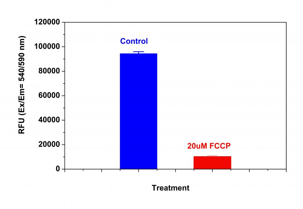
- 检测Ex / Em = 540/590 nm(截止= 570 nm)的荧光增加(底部读取模式)
溶液配制
工作溶液配制
将50 µL的200X MitoTell 橙色(组分A)添加到10 mL的测定缓冲液A(组分B)中,并充分混合以制成MitoTell Orange工作溶液,避光。
实验步骤
1.用测试化合物处理细胞一段时间,以诱导细胞凋亡,并建立平行对照实验。
阴性对照:仅用载体处理细胞。
阳性对照:在37°C 5%CO2培养箱中,以5-50 µM的浓度用FCCP或CCCP处理细胞15至30分钟。 注意:CCCP或FCCP可以与MitoTell Orange同时添加。 为了获得最佳结果,可能需要为每个单独的细胞系滴定CCCP或FCCP。
2.取出细胞培养基。注意:在添加MitoTell Orange工作溶液之前,必须除去细胞培养基。
3.将100 µL /孔/ 96孔板或25 µL /孔/ 384孔板的MitoTell Orange工作溶液添加到细胞板中。
4.将板在5%CO2、37°C的培养箱中避光放置15-30分钟。注意:适当的孵育时间取决于所用的单个细胞类型和细胞浓度。优化每个实验的孵育时间。
5.将50 µL /孔/ 96孔板或12.5 µL /孔/ 384孔板的测定缓冲液B(组分C)添加到细胞板中。注意:加载后请勿洗涤细胞。对于非贴壁细胞,建议在加入测定缓冲液B(组分C)后,以800 rpm离心细胞板2分钟,然后制动。
6.在加入分析缓冲液B(成分C)10至30分钟后,使用荧光酶标仪(底部读取模式)在Ex/Em=640/680 nm(截止=665 nm)处检测荧光强度,可以使用终点模式,也可以使用动力学模式。
参考文献
Safranine O as a fluorescent probe for mitochondrial membrane potential studied on the single particle level and in suspension
Authors: Perevoshchikova IV, Sorochkina AI, Zorov DB, Antonenko YN.
Journal: Biochemistry (Mosc) (2009): 663
Computer-assisted live cell analysis of mitochondrial membrane potential, morphology and calcium handling
Authors: Koopman WJ, Distelmaier F, Esseling JJ, Smeitink JA, Willems PH.
Journal: Methods (2008): 304
Determination of high mitochondrial membrane potential in spermatozoa loaded with the mitochondrial probe 5,5′,6,6′-tetrachloro-1,1′,3,3′-tetraethylbenzimidazolyl-carbocyanine iodide (JC-1) by using fluorescence-activated flow cytometry
Authors: Guthrie HD, Welch GR.
Journal: Methods Mol Biol (2008): 89
Effects of eprosartan on mitochondrial membrane potential and H2O2 levels in leucocytes in hypertension
Authors: Labios M, Martinez M, Gabriel F, Guiral V, Ruiz-Aja S, Beltran B, Munoz A.
Journal: J Hum Hypertens (2008): 493
Evaluation of sperm mitochondrial membrane potential by JC-1 fluorescent staining and flow cytometry
Authors: Xia XY, Wu YM, Hou BS, Yang B, Pan LJ, Shi YC, Jin BF, Shao Y, Cui YX, Huang YF.
Journal: Zhonghua Nan Ke Xue (2008): 135
How DASPMI reveals mitochondrial membrane potential: fluorescence decay kinetics and steady-state anisotropy in living cells
Authors: Ramadass R, Bereiter-Hahn J.
Journal: Biophys J (2008): 4068
Life cell quantification of mitochondrial membrane potential at the single organelle level
Authors: Distelmaier F, Koopman WJ, Testa ER, de Jong AS, Swarts HG, Mayatepek E, Smeitink JA, Willems PH.
Journal: Cytometry A (2008): 129
Mitochondrial membrane potential in axons increases with local nerve growth factor or semaphorin signaling
Authors: Verburg J, Hollenbeck PJ.
Journal: J Neurosci (2008): 8306
The mitochondrial membrane potential and Ca2+ oscillations in smooth muscle
Authors: Chalmers S, McCarron JG.
Journal: J Cell Sci (2008): 75
Cyclosporin A-induced oxidative stress is not the consequence of an increase in mitochondrial membrane potential
Authors: van der Toorn M, Kauffman HF, van der Deen M, Slebos DJ, Koeter GH, Gans RO, Bakker SJ.
Journal: Febs J (2007): 3003