上海金畔生物科技有限公司代理AAT Bioquest荧光染料全线产品,欢迎访问AAT Bioquest荧光染料官网了解更多信息。
核紫 DCS1 死细胞染料
 |
货号 | 17549 | 存储条件 | 在零下15度以下保存, 避免光照 |
| 规格 | 0.5 ml | 价格 | 1140 | |
| Ex (nm) | 371 | Em (nm) | 454 | |
| 分子量 | 744.77 | 溶剂 | DMSO | |
| 产品详细介绍 | ||||
简要概述
产品基本信息
货号:17549
产品名称:核紫 DCS1 死细胞染料
规格:0.5ml
储存条件:-20℃干燥
产品物理化学光谱特性
溶剂:DMSO
激发波长(nm):371
发射波长(nm):454
产品介绍
我们的Nuclear Violet DCS1是一种非荧光,DNA选择性和细胞不透性的紫色荧光染料,用于分析死细胞中的DNA含量。 核紫色DCS1在与双链DNA结合后具有明显增强的荧光。 它可用于荧光成像,微孔板和流式细胞仪应用。 这种DNA结合染料可用于死细胞的多色分析。
点击查看光谱
适用仪器
| 荧光显微镜 | |
| 激发: | DAPI |
| 发射: | DAPI |
| 推荐孔板: | 黑色透明孔板 |
产品说明书
染色细胞分析方案
注意:以下方案适用于大多数细胞类型。 生长培养基,细胞密度,其他细胞类型和因子的存在可能影响染色。 玻璃器皿上的残留洗涤剂也可能影响许多生物的染色,并导致在有或没有细胞存在的溶液中出现明亮染色的物质。
工作溶液配制
使用缓冲液将核紫 DCS1储备溶液(5mM)稀释到0.5-5μm的浓度。
操作步骤
1.将Nuclear Violet DCS1工作溶液加到固定、死亡或凋亡的细胞(悬浮液或粘附细胞)中,并将细胞染色15至60分钟。 在最初的实验中,最好尝试几种染料浓度来确定产生所需结果的最佳浓度。 高染料浓度倾向于引起其他细胞结构的非特异性染色。
2.用荧光显微镜、荧光酶标仪或流式细胞仪直接检测染色细胞。
图示

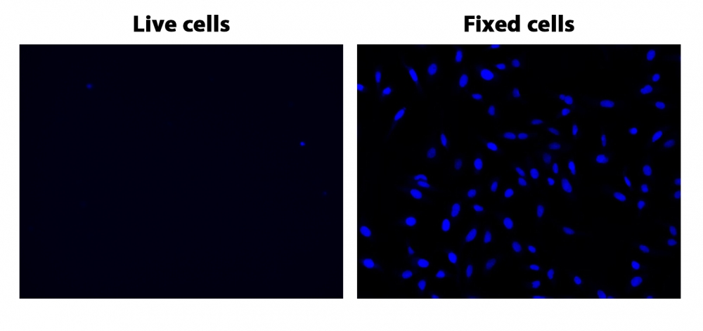
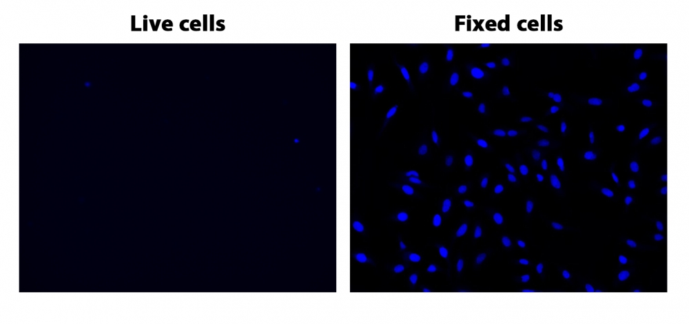
图1.在96孔板上染色的固定和活(非固定)Hela细胞,与核蓝 DCS1 1μm孵育20分钟并在DAPI通道成像。
参考文献
Assessment of laser-induced thermal damage in fresh skin with ex vivo confocal microscopy.
Authors: Ortner, Vinzent Kevin and Sahu, Aditi and Haedersdal, Merete and Rajadhyaksha, Milind and Rossi, Anthony Mario
Journal: Journal of the American Academy of Dermatology (2021): e19-e21
Development of a novel flow cytometry-based approach for reticulocytes micronucleus test in rat peripheral blood.
Authors: Chen, Yiyi and Huo, Jiao and Liu, Yunjie and Zeng, Zhu and Zhu, Xuejiao and Chen, Xuxi and Wu, Rui and Zhang, Lishi and Chen, Jinyao
Journal: Journal of applied toxicology : JAT (2021): 595-606
Exploring the utility of Deep Red Anthraquinone 5 for digital staining of ex vivo confocal micrographs of optically sectioned skin.
Authors: Ortner, Vinzent Kevin and Sahu, Aditi and Cordova, Miguel and Kose, Kivanc and Aleissa, Saud and Alessi-Fox, Christi and Haedersdal, Merete and Rajadhyaksha, Milind and Rossi, Anthony Mario
Journal: Journal of biophotonics (2021): e202000207
A novel method to purify neutrophils enables functional analysis of zebrafish hematopoiesis.
Authors: Konno, Katsuhiro and Kulkeaw, Kasem and Sasada, Manabu and Nii, Takenobu and Kaneyuki, Ayako and Ishitani, Tohru and Arai, Fumio and Sugiyama, Daisuke
Journal: Genes to cells : devoted to molecular & cellular mechanisms (2020): 770-781
Analysis of erythroid maturation in the nonlysed bone marrow with help of radar plots facilitates detection of flow cytometric aberrations in myelodysplastic syndromes.
Authors: Violidaki, Despoina and Axler, Olof and Jafari, Katayoon and Bild, Filippa and Nilsson, Lars and Mazur, Joanna and Ehinger, Mats and Porwit, Anna
Journal: Cytometry. Part B, Clinical cytometry (2020): 399-411
Identification of Cancer-Associated Circulating Cells in Anal Cancer Patients.
Authors: Carter, Thomas J and Jeyaneethi, Jeyarooban and Kumar, Juhi and Karteris, Emmanouil and Glynne-Jones, Rob and Hall, Marcia
Journal: Cancers (2020)
Influence of Polymer Charge on the Localization and Dark- and Photo-Induced Toxicity of a Potential Type I Photosensitizer in Cancer Cell Models.
Authors: Lindgren, Mikael and Gederaas, Odrun A and Siksjø, Monica and Hansen, Tom A and Chen, Lena and Mettra, Bastien and Andraud, Chantal and Monnereau, Cyrille
Journal: Molecules (Basel, Switzerland) (2020)
TEM observation of compacted DNA of Synechococcus elongatus PCC 7942 using DRAQ5 labelling with DAB photooxidation and osmium black.
Authors: Ghosh, Ilika and Atsuzawa, Kimie and Arai, Aoi and Ohmukai, Ryuzo and Kaneko, Yasuko
Journal: Microscopy (Oxford, England) (2020)
[The Establishment of a Three-color Flow Cytometry Approach for the Scoring of Micronucleated Reticulocytes in Rat Bone Marrow].
Authors: Zeng, Zhu and Zhu, Xue-Jiao and Huo, Jiao and Liu, Yun-Jie and Peng, Zi-Hao and Chen, Jin-Yao and Zhang, Li-Shi
Journal: Sichuan da xue xue bao. Yi xue ban = Journal of Sichuan University. Medical science edition (2020): 67-73
Automated Triage Radiation Biodosimetry: Integrating Imaging Flow Cytometry with High-Throughput Robotics to Perform the Cytokinesis-Block Micronucleus Assay.
Authors: Wang, Qi and Rodrigues, Matthew A and Repin, Mikhail and Pampou, Sergey and Beaton-Green, Lindsay A and Perrier, Jay and Garty, Guy and Brenner, David J and Turner, Helen C and Wilkins, Ruth C
Journal: Radiation research (2019): 342-351
相关产品
| 产品名称 | 货号 |
| 核绿 LCS1 活细胞染料 | Cat#17540 |
| 核红 LCS1 活细胞染料 | Cat#17542 |
| 核紫 LCS1 活细胞染料 | Cat#17543 |
