
上海金畔生物科技有限公司代理AAT Bioquest荧光染料全线产品,欢迎访问AAT Bioquest荧光染料官网了解更多信息。
Cell Meter 细胞衰老活性测定试剂盒
 |
货号 | 23005 | 存储条件 | 在零下15度以下保存, 避免光照 |
| 规格 | 100 Tests | 价格 | 3840 | |
| Ex (nm) | 498 | Em (nm) | 517 | |
| 分子量 | 溶剂 | |||
| 产品详细介绍 | ||||
简要概述
Cell Meter 细胞衰老活性测定试剂盒是美国AAT Bioquest生产的用于检测细胞衰老的试剂盒,细胞衰老是触发不可逆的生长停滞,以防止DNA损伤的细胞生长。与衰老相关的β-半乳糖苷酶(SA-β-gal)在衰老细胞中高度过表达,并且已被广泛用作衰老标记。 X-gal染色是一种比色法,可广泛用于检测衰老细胞中的SA-beta-gal。彩色方法有一些局限性,例如由于X-gal的细胞渗透性低,需要较长的染色时间和较低的灵敏度而需要固定样品。 Cell Meter 细胞衰老活性测定试剂盒使用Xite β-D-吡喃半乳糖苷,这是一种荧光β-Gal底物,易于进入活细胞,并被细胞内的SA-β-gal裂解,产生强烈的绿色荧光。与不渗透细胞的X-Gal底物不同,它具有出色的细胞渗透性。 Cell Meter 细胞衰老活性测定试剂盒使用户能够以更高的灵敏度和强大的性能检测衰老。 Xite产品可以很好地保留在细胞内,为荧光成像和流式细胞仪分析产生稳定的信号。金畔生物是AAT Bioquest的中国代理商,为您提供最优质的Cell Meter 细胞衰老活性测定试剂盒。
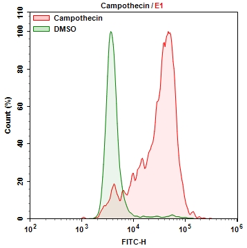
点击查看光谱
适用仪器
| 流式细胞仪 | |
| 激发: | 488nm激光 |
| 发射: | 530/30nm滤波片 |
| 通道: | FITC通道 |
| 荧光显微镜 | |
| 激发: | FITC滤波片 |
| 发射: | FITC滤波片 |
| 推荐孔板: | 黑色透明 |
产品说明书
样品实验方案
简要概述
1.根据需要处理样品。
2.准备Xite β-D-吡喃半乳糖苷工作溶液并将其添加到样品中。
3.在37°C下孵育样品15至45分钟。
4.使用FITC滤光片组监控荧光强度。
溶液制备
1.储备溶液
所有未使用的储备溶液应分为一次性使用的等分试样,并在制备后储存在-20°C下。 避免重复冻融循环。
Xite β-D-吡喃半乳糖苷储备溶液(100X):
将100 uL DMSO(组分C)添加到Xite β-D-吡喃半乳糖苷(组分A)中并充分混合。 注意:将未使用的Xite β-D-吡喃半乳糖苷储备溶液以单次使用的等分试样保存在-20°C下。
2.工作溶液
Xite β-D-吡喃半乳糖苷工作溶液(1X):
用1 mL测定缓冲液稀释10 uL Xite β-D-吡喃半乳糖苷储备溶液(100X),制成Xite β-D-吡喃半乳糖苷工作溶液(1X)。注意:应立即使用Xite β-D-吡喃半乳糖苷工作溶液。
样品操作及实验分析
以下方案可用作指导,具体实验应根据需要进行调整。
1.根据需要处理样品。
2.用您选择的缓冲液(例如DPBS)洗涤细胞。
3.加入100 uL Xite β-D-吡喃半乳糖苷工作溶液15-45分钟,然后在37°C的培养箱中孵育样品。 注意:孵育的最佳时间需要仔细确定。
4.取出工作溶液并用您选择的缓冲液洗涤细胞。
5.将细胞重悬于测定缓冲液(组分B)中,并使用流式细胞仪或带有FITC过滤器的荧光显微镜监控荧光强度。
参考文献
Cell senescence, apoptosis and DNA damage cooperate in the remodeling processes accounting for heart morphogenesis
Authors: C. I. Lorda-Diez
Journal: J Anat (2019): 815-829
Dynamic transcriptome profiling in DNA damage-induced cellular senescence and transient cell-cycle arrest
Authors: Z. Zhao
Journal: Genomics (2019): ersion=”1.0″ encoding=”UTF-8″ ?>23005.enlEndNote242417Zhao, Z.Dong, Q.Liu, X.Wei, L.Liu, L.Li, Y.Wang, X.Ministry of Education Key Laboratory of Bioinformatics, Center for Synthetic and System Biology, BNRist, Department of Automation, Tsinghua University
Stochastic modeling of aging cells reveals how damage accumulation, repair, and cell-division asymmetry affect clonal senescence and population fitness
Authors: R. Song
Journal: BMC Bioinformatics (2019): 391
Topological DNA damage, telomere attrition and T cell senescence during chronic viral infections
Authors: Y. Ji
Journal: Immun Ageing (2019): 12
Leucine reduces the proliferation of MC3T3-E1 cells through DNA damage and cell senescence
Authors: R. da Luz Dias
Journal: Toxicol In Vitro (2018): 1-10
Regulatory T cells trigger effector T cell DNA damage and senescence caused by metabolic competition
Authors: X. Liu
Journal: Nat Commun (2018): 249
The Novel Small Molecule STK899704 Promotes Senescence of the Human A549 NSCLC Cells by Inducing DNA Damage Responses and Cell Cycle Arrest
Authors: C. W. Park
Journal: Front Pharmacol (2018): 163
Hydrogen Treatment Protects against Cell Death and Senescence Induced by Oxidative Damage
Authors: A. L. Han
Journal: J Microbiol Biotechnol (2017): 365-371
MicroRNA-16 feedback loop with p53 and Wip1 can regulate cell fate determination between apoptosis and senescence in DNA damage response
Authors: M. V. C. Issler
Journal: PLoS One (2017): e0185794
Assessment of DNA Damage and Cell Senescence in Corneal Epithelial Cells Exposed to Airborne Particulate Matter (PM2.5) Collected in Guangzhou, China
Authors: Z. X. Gao
Journal: Invest Ophthalmol Vis Sci (2016): 3093-102