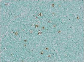
M30 Apoptosense® ELISA 细胞凋亡M30 Apoptosense ELISA试剂盒 品牌:VLVbio


品牌:VLVbio
CAS No.:
储存条件:+4℃
纯度:
产品编号

(生产商编号)

等级 规格 运输包装 零售价(RMB) 库存情况 参考值

10011

96 wells 10,000.00


* 干冰运输、大包装及大批量的产品需酌情添加运输费用


* 零售价、促销产品折扣、运输费用、库存情况、产品及包装规格可能因各种原因有所变动,恕不另行通知,确切详情请联系上海金畔生物科技有限公司。

产品描述相关资料下载相关产品浏览记录

定量检测caspase酶解的ck18片段(ccCK18/CK18F/CK18Asp396-NE/M30),不能检测坏死细胞释放出的完整的CK18。

可特异性检测小鼠模型中人的异种移植物凋亡情况。        

特异性:人                               

样品类型:血清,血浆,细胞培养上清和细胞裂解液。                                   

灵敏度:25U/L                                    

检测范围:75~1000U/L

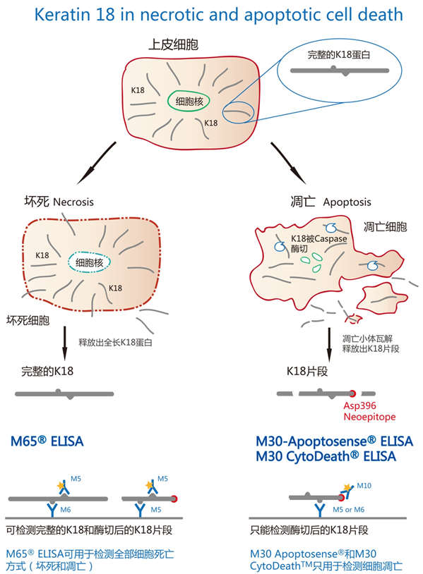
细胞角蛋白18(Cytokeratin18,简写为CK18)是上皮细胞特异性表达的一类蛋白,当上皮细胞早起发生凋亡时,具有活性的caspase(比如caspase-3,-7,9)会酶解CK18形成三个片段,其中一个片段含有neoepitope(NE),这一片段成为caspase-cleaved 

Cytokeratin18 (ccCK18),可以被单克隆抗体M30识别。

形成凋亡小体时,这些片段会释放到细胞外,当上皮细胞发生坏死时,完整的CK18被释放到细胞外。

原产品货号10010已更换为10011

M30 CytoDEATH™ ELISA M30 CytoDEATH 上皮细胞凋亡ELISA试剂盒 品牌:VLVbio


品牌:VLVbio
CAS No.:
储存条件:+4℃
纯度:
产品编号

(生产商编号)

等级 规格 运输包装 零售价(RMB) 库存情况 参考值

10900

96 wells 咨询


* 干冰运输、大包装及大批量的产品需酌情添加运输费用


* 零售价、促销产品折扣、运输费用、库存情况、产品及包装规格可能因各种原因有所变动,恕不另行通知,确切详情请联系上海金畔生物科技有限公司。

产品描述相关资料下载相关产品浏览记录

M30 CytoDeath™ ELISA可用于细胞凋亡诱导药物鉴定,包括时间进程动力学建立,剂量反应关系。也可用于大规模鉴定诱导细胞凋亡的siRNA。
该试剂盒可用于定量检测上皮细胞形成的多细胞球样体和器官培养体系的细胞凋亡M30 CytoDeath™ ELISA 采用M30抗体(检测K18上Asp396 caspase-cleavage位点) 和抗体 M6。
该抗体组合不同于M30 Apoptosense® ELISA中的抗体M30 CytoDeath™ ELISA 不能用于血清或者血浆。

GFP-CERTIFIED® Apoptosis/Necrosis detection kit 细胞凋亡/坏死检测试剂盒(荧光显微镜&流式)(GFP细胞系) 品牌:Enzo


品牌:Enzo
CAS No.:
储存条件:+4℃
纯度:
产品编号

(生产商编号)

等级 规格 运输包装 零售价(RMB) 库存情况 参考值

ENZ-51002-25

25 assays 2,600.00


* 干冰运输、大包装及大批量的产品需酌情添加运输费用


* 零售价、促销产品折扣、运输费用、库存情况、产品及包装规格可能因各种原因有所变动,恕不另行通知,确切详情请联系上海金畔生物科技有限公司。

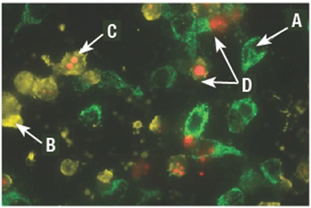
产品描述相关资料下载相关产品浏览记录 检测正常细胞、早期凋亡、晚期凋亡和坏死细胞,与GFP和其他荧光探针兼容 

应用:荧光显微镜观察 荧光检测 流式细胞仪

组分:凋亡检测试剂(Annexin V-EnzoGold)

坏死检测试剂(Red)

凋亡诱导试剂 (十字孢碱)

结合缓冲液 (10×)

规格:25次检测

Cell Meter 固定化细胞和组织TUNEL细胞凋亡测定试剂盒*绿色荧光* 货号22851-AAT Bioquest荧光染料

上海金畔生物科技有限公司代理AAT Bioquest荧光染料全线产品,欢迎访问AAT Bioquest荧光染料官网了解更多信息。

Cell Meter 固定化细胞和组织TUNEL细胞凋亡测定试剂盒*绿色荧光*

Cell Meter 固定化细胞和组织TUNEL细胞凋亡测定试剂盒*绿色荧光*

Cell Meter 固定化细胞和组织TUNEL细胞凋亡测定试剂盒*绿色荧光*    货号22851 货号 22851 存储条件 在零下15度以下保存, 避免光照
规格 25 Tests 价格 4884
Ex (nm) 498 Em (nm) 517
分子量 溶剂
产品详细介绍

简要概述

产品基本信息

货号:22851

产品名称:Cell Meter 固定化细胞和组织TUNEL细胞凋亡测定试剂盒*绿色荧光*

规格:25 Tests

储存条件:-15℃避光防潮

保质期:12个月


产品物理化学光谱特性

Ex(nm):498

Em(nm):517


适用仪器


流式细胞仪  
激发: 488nm激光
发射: 530/30nm滤波片
通道: FITC通道
荧光显微镜  
激发: FITC滤波片组
发射: FITC滤波片组
推荐孔板: 黑色透明


产品介绍

Cell Meter 固定化细胞和组织TUNEL细胞凋亡检测试剂盒提供了一种强大荧光检测工具,可方便地检测由凋亡引起的DNA片段化。该检测方法是一种非放射性,简单,准确和快速的方法,可通过对DNA片段进行成像来检测固定细胞和组织中的细胞凋亡。TUNEL分析使用末端脱氧核苷酸转移酶(TdT)催化在片段化DNA的3′-羟基末端掺入荧光素12-dUTP。通过荧光显微镜或流式细胞仪(用530/30 nm发射滤光片在488 nm激发)分析荧光素标记的DNA。该试剂盒可用于检测固定细胞和福尔马林固定的石蜡包埋的组织切片的凋亡。金畔生物是AAT Bioquest的中国代理商,为您提供最优质的Cell Meter 固定化细胞和组织TUNEL细胞凋亡测定试剂盒。 


图示


Cell Meter 固定化细胞和组织TUNEL细胞凋亡测定试剂盒*绿色荧光*    货号22851

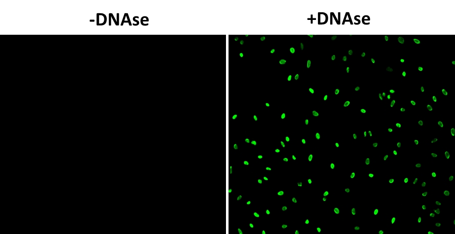
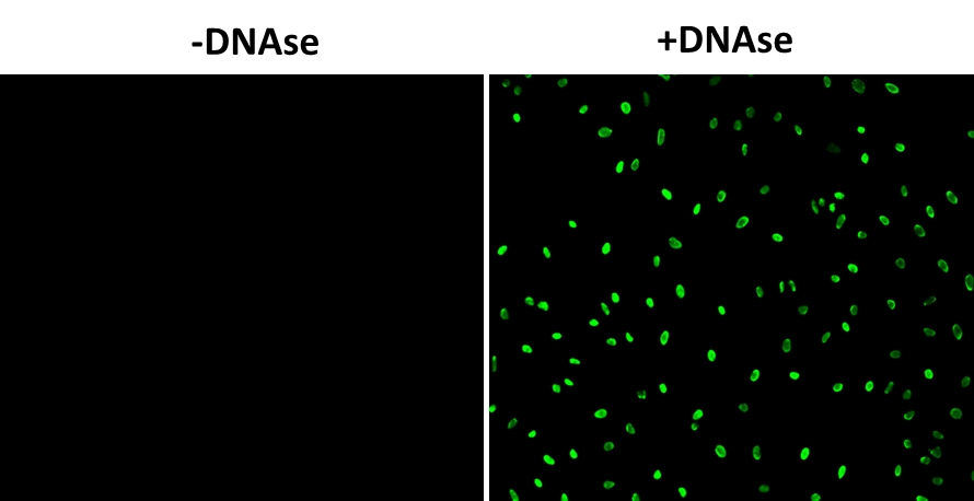
图1.固定HeLa细胞,并在37°C下用或不使用DNAse处理60分钟。然后将细胞用Cell Meter TUNEL细胞凋亡检测试剂盒染色。DNA链断裂在DNAse处理的细胞中显示出强烈的荧光染色。使用FITC滤光片组通过荧光显微镜检测信号。


参考文献

Exosomes derived from adipose tissue, bone marrow, and umbilical cord blood for cardioprotection after myocardial infarction.
Authors: Xu, Huiyu and Wang, Zhongchao and Liu, Longmei and Zhang, Baoxia and Li, Bao
Journal: Journal of cellular biochemistry (2020): 2089-2102

Improvement of in situ Follicular Activation and Early Development in Cryopreserved Human Ovarian Cortical Tissue by Co-Culturing with Mesenchymal Stem Cells.
Authors: Hosseini, Marzieh and Salehpour, Saghar and Ghaffari Novin, Marefat and Shams Mofarahe, Zahra and Abdollahifar, Mohammad-Amin and Piryaei, Abbas
Journal: Cells, tissues, organs (2020): 1-11

Brain tissue saving effects by single-dose intralesional administration of Neuroprotectin D1 on experimental focal penetrating brain injury in rats.
Authors: Berg, Rand Wilcox Vanden and Davidsson, Johan and Lidin, Erik and Angéria, Maria and Risling, Mårten and Günther, Mattias
Journal: Journal of clinical neuroscience : official journal of the Neurosurgical Society of Australasia (2019): 227-233

Cathepsin K Knockout Exacerbates Haemorrhagic Transformation Induced by Recombinant Tissue Plasminogen Activator After Focal Cerebral Ischaemia in Mice.
Authors: Zhao, Rong and He, Xin-Wei and Shi, Yan-Hui and Liu, Yi-Sheng and Liu, Feng-Di and Hu, Yue and Zhuang, Mei-Ting and Feng, Xiao-Yan and Zhao, Lei and Zhao, Bing-Qiao and Liu, Hui-Qin and Shi, Guo-Ping and Liu, Jian-Ren
Journal: Cellular and molecular neurobiology (2019): 823-831

Classification, Scoring, and Quantification of Cell Death in Tissue Sections.
Authors: Janke, Laura J and Ward, Jerrold M and Vogel, Peter
Journal: Veterinary pathology (2019): 33-38

Comparison between Slow Freezing and Vitrification for Human Ovarian Tissue Cryopreservation and Xenotransplantation.
Authors: Lee, Sanghoon and Ryu, Ki-Jin and Kim, Boram and Kang, Dahyeon and Kim, Yoon Young and Kim, Tak
Journal: International journal of molecular sciences (2019)

Correlation of MiR-152 expression with VEGF expression in placental tissue of preeclampsia rat and its influence on apoptosis of trophoblast cells.
Authors: Zhang, L and Yuan, J-M and Zhao, R-H and Wang, L-M and Tu, Z-B
Journal: European review for medical and pharmacological sciences (2019): 3553-3560

Effect of cryopreservation techniques on proliferation and apoptosis of cultured equine ovarian tissue.
Authors: Gastal, G D A and Aguiar, F L N and Ishak, G M and Cavinder, C A and Willard, S T and Ryan, P L and Feugang, J M and Gastal, E L
Journal: Theriogenology (2019): 88-94

Exosomal miR-320d derived from adipose tissue-derived MSCs inhibits apoptosis in cardiomyocytes with atrial fibrillation (AF).
Authors: Liu, Lina and Zhang, Haoran and Mao, Hongyu and Li, Xiaohong and Hu, Yamin
Journal: Artificial cells, nanomedicine, and biotechnology (2019): 3976-3984

IL-10 inhibits apoptosis in brain tissue around the hematoma after ICH by inhibiting proNGF.
Authors: Song, L and Xu, L-F and Pu, Z-X and Wang, H-H
Journal: European review for medical and pharmacological sciences (2019): 3005-3011

说明书
Cell Meter 固定化细胞和组织TUNEL细胞凋亡测定试剂盒*绿色荧光*.pdf

VLVbio 细胞凋亡坏死检测试剂盒

  • 产品特性
  • 相关资料
  • Q&A
  • 参考文献

肝病研究专家——VLVbio品牌

VLVbio 细胞凋亡坏死检测试剂盒


◆原理


来自瑞典的 VLVbio品牌,专注于研究检测细胞死亡方式(凋亡或坏死)的 ELISA 试剂盒。

上皮特异的中间丝细胞角蛋白18(keratin 18, CK18)在不同类型的细胞死亡中有不同的命运。在细胞凋亡中,CK18 被从两个位点裂解成三个片段,其中一个片段被单克隆抗体 M30 特异性识别。在细胞坏死中,CK18 是不被裂解的,释放出完整的 CK18。PEVIVA 产品主要是通过检测不同状态的 CK18 的含量来判断细胞死亡的方式。可用于肿瘤和肝病细胞凋亡检测方面的研究,提供的试剂均通过 ISO9001:2000 和 ISO13845:2003 认证。


◆优点特色


 M30 Apoptosense® ELISA 和 M30 CytoDeath™ ELISA 这两个试剂盒是利用 M30 抗体来特异性检测角蛋白18(keratin 18, CK18)片段,不能检测坏死细胞释放出的完整的 CK18。两者的不同之处是 M30 Apoptosense® ELISA 使用 mAb M5 作为捕获抗体,而 M30 CytoDeath ELISA 使用 mAb M6 作为捕获抗体。

M65® ELISA和M65 EpiDeath® ELISA 这两个试剂盒用于检测 CK18 的总量,包含细胞凋亡过程中释放的 CK18 片段和细胞坏死过程中释放的完整 CK18。这两个试剂盒常和 M30 Apoptosense® ELISA(特异性检测细胞凋亡)联用,用于判断和定量上皮细胞死亡的方式。

M65 EpiDeath® ELISA 与 M65® ELISA 不同之处是 M65 EpiDeath® ELISA 使用 mAb M5 作为捕获抗体,而 M65® ELISA 使用 mAb M6 作为捕获抗体。与 M65® ELISA 相比,M65 EpiDeath® ELISA 有更好的特异性结合,以及更低的背景。


产品名称

样品

检测线

检测范围

检测时间

检测对象

M30 Apoptosense® ELISA

细胞裂解液、细胞培养上清

人血清、人血浆

25 U/L

75-1,000 U/L

约4小时

CK18 片段

M30 CytoDeath ELISA

细胞裂解液、细胞培养上清

60 U/L

250-3,000 U/L

约4小时

CK18 片段

M65® ELISA

细胞培养上清液、人血清、人血浆

11 U/L

125-2,000 U/L

约2小时

CK18 片段和完整的CK18

M65 EpiDeath® ELISA

细胞培养上清液、人血清、人血浆

25 U/L

67-5,000 U/L

约4小时

CK18 片段和完整的CK18


VLVbio 细胞凋亡坏死检测试剂盒


◆案例应用


(1)M5 Keratin 18 antibody

可以识别 CK18 片段和完整的 CK18。

应用:WB,ICC,IHC(FFPE和FS),IP,流式细胞检测(FACS)


(2)M6 Keratin 18 antibody

可以识别 CK18 片段和完整的 CK18。

应用:WB,ICC,IHC(FFPE和FS),IP,流式细胞检测(FACS)


产品信息:


产品编

品名

包装

10011

M30 Apoptosense® ELISA*

96 wells

10020

M65® ELISA*

96 wells

10040

M65 EpiDeath® ELISA

96 wells

10900

M30 CytoDeath™ ELISA

96 wells

10700

M30 CytoDEATH (unlabeled)

200 tests

10750

M30 CytoDEATH Biotin

200 tests

10800

M30 CytoDEATH Fluorescein

200 tests

10830

M30 CytoDEATH Orange

200 tests

10850

M30 CytoDEATH Red

200 tests

10600

M5 Keratin 18 antibody

200 tests

10650

M6 Keratin 18 antibody

200 tests


*通过欧洲 CE 认证

产品编号 产品名称 产品规格 产品等级
10011 M30 Apoptosense®ELISA* 96 wells
10020 M65® ELISA* 96 wells
10040 M65 EpiDeath®ELISA 96 wells
10900 M30 CytoDeath™ ELISA 96 wells
10700 M30 CytoDEATH™ (unlabeled) 200 tests
10750 M30 CytoDEATH™ Biotin 200 tests
10800 M30 CytoDEATH™ Fluorescein 200 tests
10830 M30 CytoDEATH™ Orange 200 tests
10850 M30 CytoDEATH™Red 200 tests
10600 M5 Keratin 18 antibody 200 tests
10650 M6 Keratin 18 antibody 200 tests

​anti-Asc, pAb (AL177) 凋亡相关斑点样蛋白 Asc 检测及功能抗体

  • 产品特性
  • 相关资料
  • Q&A
  • 参考文献

​anti-Asc, pAb (AL177)                              凋亡相关斑点样蛋白 Asc 检测及功能抗体anti-Asc,pAb (AL177)

凋亡相关斑点样蛋白 Asc 检测及功能抗体


Asc 促进半胱天冬酶介导的细胞凋亡。这种促凋亡活性主要通过半胱天冬酶-9 的活性介导。它是炎症小体的一个组成部分,炎症小体是包含 NLRP3/NALP3、CARD8 和 CASP1 的蛋白复合体,其功能是激活促炎性半胱氨酸蛋白酶。


产品详情

别名

 Pycard、甲基化诱导沉默的靶点1、TMS1、Caspase Recruitment Domain-containing Protein 5、

 细胞凋亡相关Speck样蛋白 CARD、CARD5

产品类型

 多克隆抗体

免疫动物/宿主

 兔

免疫原/抗原

 对应于 N 末端人 Asc 的氨基酸合成肽.

应用

 免疫细胞化学(1:200)
 免疫组织化学(1:500;paraffin sections)
 免疫沉淀(1:200)
 Western Blot(1:1000)
 功能应用(见参考文献 [1])

交叉反应性

 人、小鼠

特异性

 识别人和小鼠 Asc.

纯度

 表位亲和纯化

浓度

 1 mg/mL

配方

 液体,溶解在含有 10% 甘油和 0.02% 叠氮钠的 PBS 中

运输

 干冰

短时间储存

 +4°C

长时间储存

 -20°C

参考文献

[1]

NALP3 forms an IL-1beta-processing inflammasome with increased activity in Muckle-Wells autoinflammatory disorder: L. Agostini,et al.; Immunity 20, 319 (2004)


[2]

P2X7 Receptor Differentially Couples to Distinct Release Pathways for IL-1beta in Mouse Macrophage: P. Pelegrin, et al.; J. Immunol. 180, 7147 (2008)


[3]

Inflammatory role of ASC in antigen-induced arthritis is independent of caspase-1, NALP-3, and IPAF: L. Kolly, et al.; J. Immunol. 183, 4003 (2009)


[4]

Activation of autophagy by inflammatory signals limits IL-1b production by targeting ubiquitinatedinflammasomes for destruction: C.-S. Shi, et al.; Nat. Immunol. 13, 255  (2012)


[5]

NLRP3 is activated in Alzheimer's disease and contributes to pathology in APP/PS1 mice: M.T. Heneka, et al.;  Nature 493, 674 (2013)


[6]

Promyelocytic leukemia protein interacts with the apoptosis-associated speck-like protein to limit inflammasome activation: J.K. Dowling, et al.; J. Biol. Chem. 289, 6429 (2014)


[7]

Localization and functionality of the inflammasome in neutrophils: M. Bakele, et al.; J. Biol. Chem. 289, 5320 (2014)


[8]

The adaptor ASC has extracellular and 'prionoid' activities that propagate inflammation: B.S. Franklin, et al.; Nat. Immunol. 15, 727 (2014)


[9]

The NLRP3 inflammasome is released as a particulate danger signal that amplifies the inflammatory response: A. Baroja-Mazo, et al.; Nat. Immunol. 15, 738 (2014)


[10]

Aging-associated TNF production primes inflammasome activation and Nlrp3-related metabolic disturbances: F. Bauernfeind, et al.; J. Immunol. 197, 2900 (2016)


[11]

An Investigation of Inflammatory and Metabolic Blood-Based Biomarkers of Cognitive Decline: H. Wolfe; Thesis Univ. Dublin, Trinity College Dep. of Physiol. (2018)


产品编号 产品名称 产品规格 产品等级
AG-25B-0006-C100 anti-Asc, pAb (AL177)
 Asc多抗(AL177)
100 μg

GFP-CERTIFIED® 细胞凋亡/坏死检测试剂盒 GFP-CERTIFIED® Apoptosis/Necrosis detection kit

  • 产品特性
  • 相关资料
  • Q&A
  • 参考文献

GFP-CERTIFIED® 细胞凋亡/坏死检测试剂盒GFP-CERTIFIED® 细胞凋亡/坏死检测试剂盒                              GFP-CERTIFIED® Apoptosis/Necrosis detection kit

GFP-CERTIFIED® Apoptosis/Necrosis detection kit

可进行区分正常细胞、早期凋亡细胞、晚期凋亡细胞和坏死细胞的多重检测,可与 GFP 和其他绿色荧光探针兼容。


● 可以兼容 GFP 和其他绿色荧光探针

● 容易区分正常细胞、早期凋亡细胞、晚期凋亡细胞和坏死细胞

● 已根据荧光显微镜和流式细胞仪等应用进行过优化

● 适用于死亡途径分析及药物、毒素研究


从凋亡到坏死的过渡是一个松散定义的连续体。质膜细胞外表面上磷脂酰丝氨酸(PS)的暴露仍是早期细胞凋亡的共同标志。这时候在Ca2+存在下,磷脂结合蛋白例如 Annexin V 与 PS 具有亲和力。由于 Annexin V 不具有细胞渗透性,所以这种胞外PS的结合是早期凋亡细胞对具有选择性的。

同样,质膜完整性的丧失,如不具有膜渗透性DNA嵌入染料若能标记出细胞核,则是表明晚期细胞凋亡和坏死的直接方法。考虑到Annexin V通常与荧光素(又称FITC)偶联,检测绿色荧光蛋白(GFP)表达细胞中的凋亡时可能会出现问题。以上两个荧光团的发射图谱会发生光谱重叠,使信号之间的区分变得困难或不可能,从而损害了总体上的数据质量。GFP-CERTIFIED® 细胞凋亡/坏死检测系统专为表达GFP的细胞系以及表达蓝色或青色荧光蛋白(BFP、CFPs)的细胞而设计,同时也可以应用于不带荧光蛋白的细胞的凋亡和坏死的细胞。另外,本试剂盒适合用于活细胞或结合了探针(如标记抗体、或与荧光素或香豆素显示相似光谱特性的其他荧光偶联物)的细胞。本试剂盒包含了用于确定细胞凋亡及坏死的早期和晚期阶段中所需的全部试剂。Annexin V -EnzoGold(增强型Cyanine-3)偶联物能够检测不同于荧光素或GFP的细胞凋亡。坏死检测试剂(红色)类似于发出红色的染料7-AAD,可促进晚期细胞凋亡和坏死检测。本试剂盒还包含了可用作阳性对照的凋亡诱导剂(Staurosporine)。检测试剂可单独使用或分别与其他单独或多重应用组合使用。本试剂盒还可以通过流式细胞仪和荧光/共焦显微镜提供快速、准确的检查结果。

GFP-CERTIFIED® 细胞凋亡/坏死检测试剂盒                              GFP-CERTIFIED® Apoptosis/Necrosis detection kit

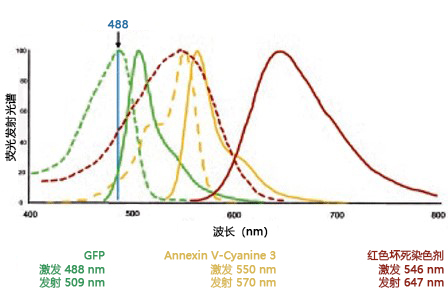
GFP、Annexin V-Cyanine 3 偶联物和坏死检测试剂(红色)的激发(阴影线)和发光(固体)光谱。

三种染料都可以轻易地被 488 nm 激光源激发。所有荧光团之间的发射峰值容易区分。

GFP-CERTIFIED® 细胞凋亡/坏死检测试剂盒                              GFP-CERTIFIED® Apoptosis/Necrosis detection kit

GFP-CERTIFIED® 细胞凋亡/坏死检测试剂盒(ENZ-51002


GFP-CERTIFIED® 细胞凋亡/坏死检测试剂盒                              GFP-CERTIFIED® Apoptosis/Necrosis detection kit


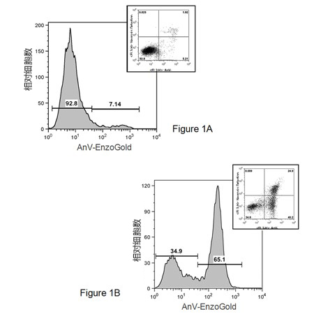
流式细胞仪。用0.2%DMSO(panel A)模拟诱导 Jurkat 细胞,或用 2 μM 星形孢菌素(panel B)在37°C下诱导 Jurkat 细胞4h。经过处理后,在含有 Annexin V-Cyanine 3 和坏死检测试剂(红色;一种远红色发光的 DNA 嵌入染料)的缓冲液中孵育细胞,然后用 488 nm 激光的流式细胞仪进行分析,用 FL2(细胞凋亡)和 FL3(坏死)通道进行荧光检测。

模拟诱导的细胞凋亡和坏死主要呈阴性。处理4h 后,会出现3种细胞群:

(1)可以存活且不凋亡、不坏死的细胞(Annexin V-Cyanine 3、坏死检测试剂呈阴性);

(2)正在经历凋亡的细胞(Annexin V-Cyanine 3 呈阳性和 NDR 呈阴性);

(3)处于晚期凋亡和早期坏死的细胞(Annexin V-Cyanine 3 和坏死检测试剂呈阳性)。


GFP-CERTIFIED® 细胞凋亡/坏死检测试剂盒                              GFP-CERTIFIED® Apoptosis/Necrosis detection kit

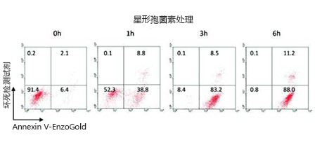
用 2.5 µM 星形孢菌素刺激 Jurkat 细胞 0-6 h。使用 Jurkat 悬浮细胞的流式细胞仪结果表现为早期凋亡(Annexin V-EnzoGold)和晚期凋亡/坏死(Annexin V-EnzoGold 和坏死染色)。


◆产品规格


 应用:

 流式细胞仪、荧光显微镜、荧光检测。

 使用事项/稳定性:

 Annexin V-EnzoGold 和结合缓冲液应在4°C储存。
 其他全部试剂应在-20°C储存。
 重组诱导剂(星形孢菌素)应在-20°C下储存。具体请参阅说明书。

 储存条件:

 避光。

 运输:

 蓝冰运输。

 短期储存温度:

 4°C

 试剂盒构成:

 凋亡检测试剂 (Annexin   V-EnzoGold)
 坏死检测试剂
 凋亡诱导剂(Staurosporine)
 结合缓冲液(10X)

 技术信息/产品说明:

 应用说明:
 Image-Based Analysis of a Human Neurosphere Stem Cell Model for the Evaluation of Potential 

 Neurotoxicants
 引用样本:
 For an overview on cited samples please click here.

 监管状况:

 RUO-仅供研究用


点击下载此处下载相关产品宣传页

 其他自噬相关产品


  自噬抑制剂


产品编号

产品名称

应用

规格

BML-AP502-0025

3-Methyladenine

3-甲基腺嘌呤,PI3激酶抑制剂

25 mg

BML-CM110-0100

Bafilomycin A1

巴伐洛霉素,ATPase抑制剂

100 μg


自噬激活剂


产品编号

产品名称

应用

规格

产品编号

产品名称

BML-PE180-0001

Thapsigargin

毒胡萝卜素,ATPase抑制剂

1 mg

BML-PE180-0001

Thapsigargin

BML-CC104-0010

Tunicamycin

衣霉素,GlcNAc磷酸转移酶抑制剂

10 mg

BML-CC104-0010

Tunicamycin

ALX-550-084-G005

Carbamazepine

卡马西平,镇痉剂和止痛剂

5 g

ALX-550-084-G005

Carbamazepine

BML-SL100-0005

C2 Ceramide

C2-神经酰胺,细胞渗透性神经酰胺类似

5 mg

BML-SL100-0005

C2 Ceramide

BML-A275-0005

Rapamycin

雷帕霉素,免疫抑制剂

5 mg

BML-A275-0005

Rapamycin

BML-CA409-0050

Xestospongin C

IP3受体抑制剂

50 μg

BML-CA409-0050

Xestospongin C


自噬化合物库 


产品编号

产品名称

应用

规格

BML-AP502-0025

3-Methyladenine

3-甲基腺嘌呤,PI3激酶抑制剂

25 mg

BML-CM110-0100

Bafilomycin A1

巴伐洛霉素,ATPase抑制剂

100 μg

参考文献


 1.

Inhibitory role of TRIP-Br1/XIAP in necroptosis under nutrient/serum starvation: Z. Sandag, et al.; Mol. Cells 43, 236 (2020), 摘要全文

 2.

Inhibitory role of AMP‑activated protein kinase in necroptosis of HCT116 colon cancer cells with p53 null mutation under nutrient starvation: D.T. Le, et al.; Int. J. Oncol. 54, 702 (2019), 摘要;


 3.

Injectable and Quadruple-Functional Hydrogel as an Alternative to Intravenous Delivery for Enhanced Tumor Targeting: Z.Q. Zhang, et al.; ACS Appl. Mater. Interfaces 11, 34634 (2019), 摘要;


 4.

Intrinsic antibacterial activity of nanoparticles made of β-cyclodextrins potentiates their effect as drug nanocarriers against tuberculosis: A. Machelart, et al.; ACS Nano 13, 3992 (2019), 摘要;


 5.

Knockdown of TM9SF4 boosts ER stress to trigger cell death of chemoresistant breast cancer cells: Y. Zhu, et al.; Oncogene 38, 5778 (2019), Application(s): Flow cytometry using MCF-7 cells, 摘要;


  6.

PKM2 coordinates glycolysis with mitochondrial fusion and oxidative phosphorylation: T. Li, et al.; Protein Cell 10, 583 (2019), Application(s): Flow cytometry using H1299 and HepG2 cells, 摘要;


 7.

Tamoxifen acts on Trypanosoma cruzi sphingolipid pathway triggering an apoptotic death process: M. Landoni, et al.; Biochem. Biophys. Res. Commun. 516, 934 (2019), 摘要;


 8.

Anti-leukemic efficacy of BET inhibitor in a preclinical mouse model of MLL-AF4+ infant ALL: M. Bardini, et al.; Mol. Cancer Ther. 17, 1705 (2018), 摘要;


 9.

Pivotal role of human stearoyl-CoA desaturases (SCD1 and 5) in breast cancer progression: oleic acid-based effect of SCD1 on cell migration and a novel pro-cell survival role for SCD5: C. Angelucci, et al.; Oncotarget. 9, 24364 (2018), Application(s): Fluorescent microscopy with MCF-7 cells, 摘要全文


10.

Preclinical efficacy and safety of CD19CAR cytokine-induced killer cells transfected with Sleeping Beauty transposon for the treatment of acute lymphoblastic leukemia: C.F. Magnani, et al.; Hum. Gene. Ther. 29, 605 (2018), 摘要;


11.

Protective effect of a newly developed fucose-deficient recombinant antithrombin against histone-induced endothelial damage: T. Iba, et al.; Int. J. Hematol. 107, 528 (2018), 摘要;


12.

The poly (ADP-ribose) polymerase inhibitor olaparib induces up-regulation of death receptors in primary acute myeloid leukemia blasts by NF-κB activation: I. Faraoni, et al.; Cancer Lett. 423, 127 (2018), Application(s): Flow cytometry with primary AML cells, 摘要;


13.

Toxicity and phototoxicity in human ARPE-19 retinal pigment epithelium cells of dyes commonly used in retinal surgery: D. Awad, et al.; Eur. J. Ophthalmol. 28, 433 (2018), 摘要全文


14.

Cluster microRNAs miR‐194 and miR‐215 suppress the tumorigenicity of intestinal tumor organoids: T. Nakaoka, et al.; Cancer Sci. 108, 678 (2017), Application(s): Flow cytometry using organoid culture of mouse intestinal tumors, 摘要

15.

Elaidic Acid, a Trans-Fatty Acid, Enhances the Metastasis of Colorectal Cancer Cells: H. Ohmori, et al.; Pathobiology 84, 144 (2017), 摘要;


16.

Metabolic modulation of Ewing sarcoma cells inhibits tumor growth and stem cell properties: A. Dasgupta, et al.; Oncotarget. 8, 77292 (2017), Application(s): Flow cytometry with A673 cells, 摘要全文


17.

Multiple hyperthermia-mediated release of TRAIL/SPION nanocomplex from thermosensitive polymeric hydrogels for combination cancer therapy: Z.Q. Zhang, et al.; Biomaterials. 132, 16 (2017), Application(s): Confocal microscopy with PC3 and U-87 MG cells, 摘要;


18.

Pro-metastatic intracellular signaling of the elaidic trans fatty acid: K. Fujii, et al.; Int. J. Oncol. 50, 85 (2017), 摘要;


19.

Visualization of ceramide channels in lysosomes following endogenous palmitoyl-ceramide accumulation as an initial step in the induction of necrosis: M. Yamane, et al.; Biochem. Biophys. Rep. 11, 174 (2017), Application(s): Confocal microscopy with A549 cells, 摘要全文


20.

Immunotherapy of acute leukemia by chimeric antigen receptor-modified lymphocytes using an improved Sleeping Beauty transposon platform: C.F. Magnani, et al.; Oncotarget 7, 51581 (2016), Application(s): Flow cytometry using KG-1, REH, Nalm-6, and THP-1 cells, 摘要全文


21.

Novel long chain fatty acid derivatives of quercetin-3-O-glucoside reduce cytotoxicity induced by cigarette smoke toxicants in human fetal lung fibroblasts: N. Sumudu, et al.; Eur. J. Pharmacol. 781, 128 (2016), 摘要;


22.

Phlorofucofuroeckol Improves Glutamate-Induced Neurotoxicity through Modulation of Oxidative Stress-Mediated Mitochondrial Dysfunction in PC12 Cells: J.J. Kim, et al.; PLoS One 11, e0163433 (2016), 摘要全文


23.

PKLR promotes colorectal cancer liver colonization through induction of glutathione synthesis: A. Nguyen, et al.; J. Clin. Invest. 126, 681 (2016), Application(s): Assessed Apoptosis, Flow cytometry, 摘要

24.

Protective effects of β‐casofensin, a bioactive peptide from bovine β‐casein, against indomethacin‐induced intestinal lesions in rats: C. Bessette, et al.; Mol. Nutr. Food Res. 60, 823 (2016), 摘要;


25.

BRCA1, PARP1 and γH2AX in acute myeloid leukemia: Role as biomarkers of response to the PARP inhibitor olaparib: I. Faraoni, et al.; Biochim. Biophys. Acta 1852, 462 (2015), Application(s): Flow cytometry using primary AML cells, 摘要;


26.

Growth-promoting and tumourigenic activity of c-Myc is suppressed by Hhex: V. Marfil, et al.; Oncogene 34, 3011 (2015), Application(s): Flow cytometry using HO15.19 and TGR-1 cells, 摘要;


27.

common set of proteins modulated in Chikungunya virus infection: R. Abraham, et al.; J. Proteomics. 120, 126 (2015), Application(s): Flow cytometry using HEK293 cells, 摘要;


28.

Identification of novel osteogenic compounds by an ex-vivo sp7: luciferase zebrafish scale assay: E. de Vrieze, et al.; Blood 74, 106 (2015), Application(s): Fluorescence microscopy using fish scales, 摘要;


29.

Impaired oxidative phosphorylation regulates necroptosis in human lung epithelial cells: M.J. Koo, et al.; Biochem. Biophys. Res. Commun. 464, 875 (2015), Application(s): Apoptosis/necrosis assay, 摘要;


30.

Influenza virus M2 targets cystic fibrosis transmembrane conductance regulator for lysosomal degradation during viral infection: J.D. Londino, et al.; FASEB J. 29, 2712 (2015), Application(s): Flow cytometry using HEK293 cells, 摘要

31.

Inhibition of autophagy exerts anti-colon cancer effects via apoptosis induced by p53 activation and ER stress: K. Sakitani, et al.; BMC Cancer 15, 795 (2015), Application(s): Flow cytometric analysis of apoptosis, 摘要全文


32.

N-terminal functional domain of Gasdermin A3 regulates mitochondrial homeostasis via mitochondrial targeting: P.H. Lin, et al.; J. Biomed. Sci. 22, 44 (2015), Application(s): Fluorescence microscopy using HaCaT and HEK293 cells, 摘要全文


33.

Novel carbocyclic curcumin analog CUR3d modulates genes involved in multiple apoptosis pathways in human hepatocellular carcinoma cells.: K.S. Bhullar, et al. ; Chem. Biol. Interact. 242, 107 (2015), Application(s): Fluorescence microscopy, 摘要;


34.

NPD1-mediated stereoselective regulation of BIRC3 expression through cREL is decisive for neural cell survival: J.M. Calandria, et al.; Cell Death Differ. 22, 1363 (2015), Application(s): Assay, 摘要;


35.

ROS-induced oxidative stress and apoptosis-like event directly affect the cell viability of cryopreserved embryogenic callus in Agapanthus praecox: D. Zhang, et al.; Plant Cell Rep. 34, 1499 (2015), 摘要;


36.

The Adhesion GPCR CD97/ADGRE5 inhibits apoptosis: C.C. Hsiao, et al.; Int. J. Biochem. Cell. Biol. 65, 197 (2015), Application(s): Flow cytometry using HT1080 cells, 摘要;


37.

The effect of plasma-derived activated protein C on leukocyte cell-death and vascular endothelial damage: T. Iba, et al.; Thromb. Res 135, 963 (2015), Application(s): Fluorescence Microscopy using leukocytes, 摘要;


38.

Accumulation of cytosolic calcium induces necroptotic cell death in human neuroblastoma: M. Nomura, et al.; Cancer Res. 74, 1056 (2014), 摘要;


39.

Apoptotic and inhibitory effects on cell proliferation of hepatocellular carcinoma HepG2 cells by methanol leaf extract of Costus speciosus: S.V. Nair, et al.; Biomed. Res. Int. 2014, Article ID 637098 (2014), Application(s): Measurement by flow cytometry, 摘要全文


40.

Combination of antithrombin and recombinant thrombomodulin modulates neutrophil cell-death and decreases circulating DAMPs levels in endotoxemic rats: T. Iba, et al.; Throm. Res. 134, 169 (2014), Application(s): Detection Kit using live cells, 摘要;


41.

Fatty acid esters of ohloridzin induce apoptosis of human liver cancer cells through altered gene expression: S.V. Nair, et al.; PLoS One 9, e107149 (2014), Application(s): Fluorescence microscopy of HepG2 cells, 摘要全文


42.

Flavonoid-enriched apple fraction AF4 induces cell cycle arrest, DNA topoisomerase II inhibition, and apoptosis in human liver cancer HepG2 cells: S. Sudan, et al.; Nutr. Cancer 66, 1237 (2014), 摘要;


43.

Quercetin-3-O-glucoside induces human DNA topoisomerase II inhibition, cell cycle arrest and apoptosis in hepatocellular carcinoma cells: S. Sudan, et al.; Anticancer Res. 34, 1691 (2014), 摘要;


44.

A light-activated NO donor attenuates anchorage independent growth of cancer cells: Important role of a cross talk between NO and other reactive oxygen species: S. Sen, et al.; Arch. Biochem. Biophys. 540, 33 (2013), Application(s): Detection Kit using live cells, 摘要;


45.

Alu Elements in ANRIL Non-Coding RNA at Chromosome 9p21 Modulate Atherogenic Cell Functions through Trans-Regulation of Gene Networks: L.M. Holdt, et al.; PLoS Genet. 9, e1003588 (2013), Application(s): Detection Kit using live cells, 摘要

46.

Analysis of protein translocation into the endoplasmic reticulum of human cells: J. Dudek, et al.; Methods Mol. Biol. 1033, 285 (2013), 摘要;


47.

Characterizing virulence-specific perturbations in the mitochondrial function of macrophages infected with Mycobacterium tuberculosis: S. Jamwal, et al.; Sci. Rep. 3, 1328 (2013), 摘要

48.

Evaluation of anticancer effects and enhanced doxorubicin cytotoxicity of xanthine derivatives using canine hemangiosarcoma cell lines: T. Motegi, et al.; Res. Vet. Sci. 95, 600 (2013), Application(s): Fluorescence microscopy using canine HAS cells, 摘要;


49.

Genista sessilifolia DC. extracts induce apoptosis across a range of cancer cell lines: P. Bontempo, et al.; Cell Prolif. 46, 183 (2013), Application(s): Detection Kit using live cells, 摘要;


50.

High mobility group box 1 released from necrotic cells enhances regrowth and metastasis of cancer cells that have survived chemotherapy: Y. Luo, et al.; Eur. J. Cancer. 49, 741 (2013), Application(s): Fluorescence microscopy using CT26 cells, 摘要;


51.

Influence of gefitinib and erlotinib on apoptosis and c-MYC expression in H23 lung cancer cells: M. Suenaga, et al.; Anticancer Res. 33, 1547 (2013), 摘要;


52.

Comparison of different suicide-gene strategies for the safety improvement of genetically manipulated T cells: V. Marin, et al.; Hum. Gene Ther. Methods 23, 376 (2012), Application(s): Flow cytometry using EBV-CTL and 293 T cells, 摘要全文


53.

Maintenance of higher H2O2 levels, and its mechanism of action to induce growth in breast cancer cells: Important roles of bioactive catalase and PP2A: S. Sen, et al.; Free Radic. Biol. Med. 53, 1541 (2012), Application(s): Detection Kit using live cells, 摘要;


54.

Mesenchymal stem cells from Shwachman–Diamond syndrome patients display normal functions and do not contribute to hematological defects: V. André, et al.; Blood Cancer J. 2, e94 (2012), Application(s): Flow cytometry using human neutrophils, 摘要

55.

Methionine excess in diet induces acute lethal hepatitis in mice lacking cystathionine γ-lyase, an animal model of cystathioninuria: H. Yamada, et al.; Free Radic. Biol. Med. 52, 1716 (2012), Application(s): Fluorescence microscopy using mouse hepatocytes, 摘要;


56.

Molecular mechanism of interleukin-2-induced mucosal homeostasis : J. Mishra, et al.; Am. J. Physiol. Cell Physiol. 302, C735 (2012), Application(s): Detection Kit using live cells, 摘要全文


57.

Serratia marcescens induces apoptotic cell death in host immune cells via a lipopolysaccharide- and flagella-dependent mechanism: K. Ishii, et al.; J. Biol. Chem. 287, 36582 (2012), Application(s): Apoptosis detected in Silkworm larvae, 摘要;


58.

Serratia marcescens Induces Apoptotic Cell Death in Host Immune Cells via a Lipopolysaccharide- and Flagella-dependent Mechanism: K. Ishii, et al.; J. Biol. Chem. 287, 36582 (2012), Application(s): Detection Kit using live cells, 摘要全文


59.

Two mutations impair the stability and function of ubiquitin-activating enzyme (E1): T. Lao, et al.; J. Cell. Physiol. 227, 1561 (2012), Application(s): Viability and induction of cell death observed by confocal microscopy, 摘要全文


60.

Critical roles of Cold Shock Domain Protein A as an endogenous angiogenesis inhibitor in skeletal muscle: Y. Saito, et al.; Antioxid. Redox. Signal. 15, 2109 (2011), 摘要;


61.

Cell Surface Externalization of Annexin A1 as a Failsafe Mechanism Preventing Inflammatory Responses during Secondary Necrosis: K.E. Blume, et al.; J. Immunol. 183, 8138 (2009), 摘要全文


62. 

Supplemental Information for: Cell Surface Externalization of Annexin A1 as a Failsafe Mechanism Preventing Inflammatory Responses during Secondary Necrosis: K.E. Blume, et al.; J. Immunol. 183, (2009), (Supplemental Information), 全文

产品编号 产品名称 产品规格 产品等级
ENZ-51002-25 GFP-CERTIFIED® Apoptosis/Necrosis detection kit
细胞凋亡/坏死检测试剂盒(荧光显微镜&流式)(GFP细胞系)
25 assays
ENZ-51002-100 GFP-CERTIFIED® Apoptosis/Necrosis detection kit
细胞凋亡/坏死检测试剂盒(荧光显微镜&流式)(GFP细胞系)
100 assays

细胞凋亡in situ检测试剂盒Wako Apoptosis in situ Detection Kit Wako

  • 产品特性
  • 相关资料
  • Q&A
  • 参考文献

细胞凋亡in situ检测试剂盒Wako                              Apoptosis in situ Detection Kit WakoTUNEL法凋亡检测试剂盒

细胞凋亡in situ检测试剂盒Wako


  本试剂盒是利用TUNEL法检测细胞凋亡细胞的试剂盒。主要试剂都已试剂盒化,可简便且快捷检测细胞凋亡细胞。


◆特点


● 主要试剂都已试剂盒化

● 短时间:约2小时操作流程

● 低背景


试剂盒内容


● Protein Digestion Enzyme・・・・・・ 1 mL

● TdT・・・・・・・・・・・・・・ ・ ・ 40 μL

● TdT Substrate Solution・・・・・ ・ ・ 4.4 mL

● 100×POD-Conjugated Antibody・ ・ ・44 μL

● DNase I・・・・・・・・・・・・・ ・・4 μL

● 10×DNase I Reaction Buffer・・・ ・ ・40 μL

※DAB试剂单独出售。


◆染色案例


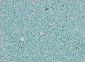
经α-Mangostin处理的小鼠移植乳腺癌(*200)


细胞凋亡in situ检测试剂盒Wako                              Apoptosis in situ Detection Kit Wako

细胞凋亡in situ检测试剂盒Wako                              Apoptosis in situ Detection Kit Wako

对照组


α-Mangostin处理组


△通过α-Mangostin处理后从抗肿瘤疗效确认癌细胞的凋亡情况。

(数据提供:大阪保健医疗大学  柴田雅朗)


产品编号 产品名称 产品规格 产品等级
293-71501 细胞凋亡in situ检测试剂盒(Wako)
Apoptosis in situ Detection Kit Wako
40次 细胞凋亡研究用
相关产品
297-71401 Apoptosis Ladder Detection Kit Wako 24 lane用 细胞凋亡研究用
293-71403 Apoptosis Ladder Detection Kit Wako 96 lane用 细胞凋亡研究用
049-22831 DAB锭(DAB•4HCl 10 mg/片)
DAB Tablet(DAB . 4HCl 10mg/Tablet)
50片 生化学用
045-22833 DAB锭(DAB•4HCl 10 mg/片)
DAB Tablet(DAB . 4HCl 10mg/Tablet)
100片 生化学用
040-27001 DAB锭(DAB•4HCl 5 mg/片)
DAB Tablet(DAB . 4HCl 5mg/Tablet)
50片 生化学用
046-27003 DAB锭(DAB•4HCl 5 mg/片)
DAB Tablet(DAB . 4HCl 5mg/Tablet)
100片 生化学用
047-27011 DAB TRIS Tablet, pH7.6 50片 生化学用