Cell Meter™ Cell Proliferation Assay Kit 货号22505-AAT Bioquest荧光染料

上海金畔生物科技有限公司代理AAT Bioquest荧光染料全线产品,欢迎访问AAT Bioquest荧光染料官网了解更多信息。

Cell Meter™ Cell Proliferation Assay Kit

Cell Meter™ Cell Proliferation Assay Kit

货号 22505 存储条件 在零下15度以下保存, 避免光照
规格 200 Tests 价格 2340
Ex (nm) 503 Em (nm) 527
分子量 N/A 溶剂
产品详细介绍

简要概述

Cell Meter BX520固定化细胞活性染料 货号22510-AAT Bioquest荧光染料

上海金畔生物科技有限公司代理AAT Bioquest荧光染料全线产品,欢迎访问AAT Bioquest荧光染料官网了解更多信息。

Cell Meter BX520固定化细胞活性染料

Cell Meter BX520固定化细胞活性染料

Cell Meter BX520固定化细胞活性染料    货号22510 货号 22510 存储条件 在零下15度以下保存, 避免光照
规格 200 Tests 价格 2412
Ex (nm) 491 Em (nm) 516
分子量 溶剂 DMSO
产品详细介绍

简要概述

产品基本信息

货号:22510

产品名称:Cell Meter BX520固色活性染料

规格:200 Tests

储存条件:保存在冰箱-15℃干燥

保质期:12个月


产品物理化学光谱特性

分子量:N/A

激发波长(nm):491

发射波长(nm):510


适用仪器


流式细胞仪  
激发: 480nm激光
发射: 530/30nm滤波片
滤波片: FITC滤波片组
荧光显微镜  
激发: FITC滤波片组
发射: FITC滤波片组
推荐孔板: 黑色透明


产品介绍 

从活细胞中区分和排除死细胞可以更清晰地分离和鉴定细胞群,Cell Meter BX520固定化细胞活性染料是细胞不可渗透的一类荧光活性染料,它们通过优化以匹配常见流式细胞仪的主要激发激光,例如350、405、488、633和647 nm。这些染料不渗入活细胞,但渗入膜受损的细胞。它们与含胺和硫醇的蛋白质以及其他细胞成分发生不可逆的反应。由于具有受损膜的死细胞或固定细胞更容易与Cell Meter BX520固定化细胞活性染料发生反应,因此比具有完整膜的活细胞的染色更亮,这些染料可用于评估哺乳动物细胞的活与死状态。使用这些染料时,需要考虑一些因素,例如,每种染料的滴定,以确保活细胞几乎没有染色。Cell Meter BX520固定化细胞活性染料通过优化,可以在488 nm的蓝色激发光中激发,并在520 nm(FITC通道)发射。与其他商业上类似的活性染料相比,这种可固定的活性染料更加耐用、稳定。金畔生物是AAT Bioquest的中国代理商,为您提供最优质的Cell Meter BX520固定化细胞活性染料。 

点击查看光谱


图示


Cell Meter BX520固定化细胞活性染料    货号22510

图1.通过Cell Meter 固定化细胞活性染料检测Jurkat细胞活性。处理Jurkat细胞并用Cell Meter BX520(Cat#22510)染色,然后固定在3.7%甲醛中并通过流式细胞仪进行分析。带有FITC通道的死细胞群体(绿色)很容易与活细胞群体(红色)区分开,在固定之前和之后都获得了几乎相同的结果。


参考文献

CFSE dilution to study human T and NK cell proliferation in vitro.
Authors: Terrén, Iñigo and Orrantia, Ane and Vitallé, Joana and Zenarruzabeitia, Olatz and Borrego, Francisco
Journal: Methods in enzymology (2020): 239-255

Using Carboxy Fluorescein Succinimidyl Ester (CFSE) to Identify Quiescent Glioblastoma Stem-Like Cells.
Authors: Azari, Hassan and Deleyrolle, Loic P and Reynolds, Brent A
Journal: Methods in molecular biology (Clifton, N.J.) (2018): 59-67

Estimates and impact of lymphocyte division parameters from CFSE data using mathematical modelling.
Authors: Mazzocco, Pauline and Bernard, Samuel and Pujo-Menjouet, Laurent
Journal: PloS one (2017): e0179768

Analysis of CFSE time-series data using division-, age- and label-structured population models.
Authors: Hross, Sabrina and Hasenauer, Jan
Journal: Bioinformatics (Oxford, England) (2016): 2321-9

Assessment of lymphocyte proliferation for diagnostic purpose: Comparison of CFSE staining, Ki-67 expression and 3H-thymidine incorporation.
Authors: Lašťovička, Jan and Rataj, Michal and Bartůňková, Jiřina
Journal: Human immunology (2016): 1215-1222

Poor association of allergen-specific antibody, T- and B-cell responses revealed with recombinant allergens and a CFSE dilution-based assay.
Authors: Eckl-Dorna, J and Campana, R and Valenta, R and Niederberger, V
Journal: Allergy (2015): 1222-9

Quality control of extracorporeal photochemotherapy: Proliferation assay using CFSE validated according to ISO 15189:2007 standards.
Authors: Faivre, Lionel and Lecouflet, Lucie and Liu, Wang-Qing and Khadher, Isabelle and Lahaie, Camille and Vidal, Michel and Legouvello, Sabine and Beaumont, Jean-Louis and Bierling, Philippe and Rouard, Hélène and Birebent, Brigitte
Journal: Cytometry. Part B, Clinical cytometry (2015): 30-9

Mathematical models for CFSE labelled lymphocyte dynamics: asymmetry and time-lag in division.
Authors: Luzyanina, Tatyana and Cupovic, Jovana and Ludewig, Burkhard and Bocharov, Gennady
Journal: Journal of mathematical biology (2014): 1547-83

Quality control of extracorporeal photochemotherapy: Proliferation assay using CFSE validated according to ISO 15189:2007 standards.
Authors: Lionel, Faivre and Lucie, Lecouflet and Wang-Qing, Liu and Isabelle, Khadher and Camille, Lahaie and Michel, Vidal and Sabine, Legouvello and Jean-Louis, Beaumont and Philippe, Bierling and Hélène, Rouard and Brigitte, Birebent
Journal: Cytometry. Part B, Clinical cytometry (2014)

Asymmetry of Cell Division in CFSE-Based Lymphocyte Proliferation Analysis.
Authors: Bocharov, Gennady and Luzyanina, Tatyana and Cupovic, Jovana and Ludewig, Burkhard
Journal: Frontiers in immunology (2013): 264

说明书
Cell Meter BX520固定化细胞活性染料.pdf

Cell Meter BX650固定化细胞活性染料 货号22520-AAT Bioquest荧光染料

上海金畔生物科技有限公司代理AAT Bioquest荧光染料全线产品,欢迎访问AAT Bioquest荧光染料官网了解更多信息。

Cell Meter BX650固定化细胞活性染料

Cell Meter BX650固定化细胞活性染料

Cell Meter BX650固定化细胞活性染料    货号22520 货号 22520 存储条件 在零下15度以下保存, 避免光照
规格 200 Tests 价格 2412
Ex (nm) 518 Em (nm) 654
分子量 844.83 溶剂 DMSO
产品详细介绍

简要概述

产品基本信息

货号:22520

产品名称:Cell Meter BX650固定化细胞活性染料

规格:200 Tests

储存条件:保存在冰箱-15℃干燥

保质期:12个月


产品物理化学光谱特性

分子量:N/A

激发波长(nm):518

发射波长(nm):654


适用仪器


流式细胞仪  
激发: 480nm激光
发射: 695/40nm滤波片
滤波片: PerCP滤波片组
荧光显微镜  
激发: TexasRed滤波片组
发射: TexasRed滤波片组
推荐孔板: 黑色透明


产品介绍 

从活细胞中区分和排除死细胞可以更清晰地分离和鉴定细胞群。Cell Meter BX650固定化细胞活性染料是一类细胞不可渗透的荧光活性染料,它们经过优化以匹配常见流式细胞仪的主要激发激光,例如350、405、488、633和647 nm。这些染料不渗入活细胞,但渗入膜受损的细胞。它们与含胺和硫醇的蛋白质以及其他细胞成分发生不可逆的反应。由于具有受损膜的死细胞或固定细胞更容易与Cell Meter BX650固定化细胞活性染料发生反应,因此比具有完整膜的活细胞的染色更亮,因此这些染料可用于评估哺乳动物细胞的活与死状态。使用这些染料时,需要考虑一些因素,例如,每种染料的滴定,以确保活细胞几乎没有染色。Cell Meter BX650固定化细胞活性染料经过优化,可以在488 nm的蓝色激光激发下以650 nm的波长发射。与其他商业上类似的活性染料相比,这种固定化细胞活性染料更加耐用、稳定。金畔生物是AAT Bioquest的中国代理商,为您提供最优质的Cell Meter BX650固定化细胞活性染料。 

点击查看光谱


图示


Cell Meter BX650固定化细胞活性染料    货号22520

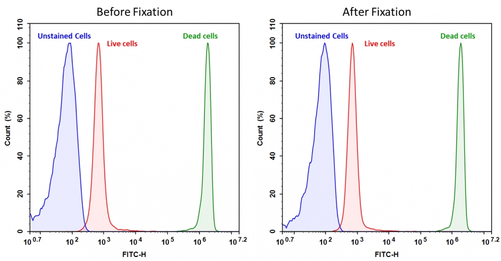
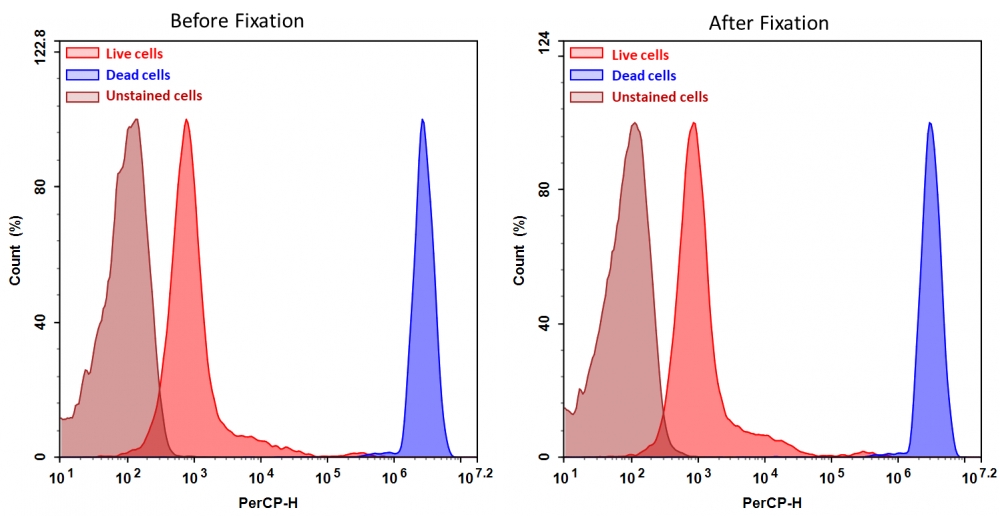
图1.通过Cell Meter 固定化细胞活性染料检测Jurkat细胞活性。处理Jurkat细胞并用Cell Meter BX650(Cat#22520)染色,然后固定在3.7%甲醛中,并通过流式细胞仪进行分析。带有PerCP通道的死细胞群体(蓝色峰)很容易与活细胞群体(红色峰)区分开,在固定之前和之后都获得了几乎相同的结果。


参考文献

Fluorescence-based method is more accurate than counting-based methods for plotting growth curves of adherent cells.
Authors: Pereira, Túlio Felipe and Levin, Gabriel and DeOcesano-Pereira, Carlos and Caodaglio, Amanda Schiersner and Fujita, André and Tonso, Aldo and Sogayar, Mari Cleide
Journal: BMC research notes (2020): 57

[Immune function of myeloid-derived suppressor cells and its mechanism in obstructive sleep apnea syndrome].
Authors: Chen, S W and Li, J and Xiang, B and Xu, S J and Wang, L
Journal: Zhonghua yi xue za zhi (2020): 295-300

CFDA-SE Combined with MACSiBeads™ Particles to Evaluate the Inhibitory Effect of Treg Cells in vitro.
Authors: Ren, Qingqi and Jiang, Chunlin and Liu, Jikui
Journal: Annals of clinical and laboratory science (2019): 740-747

Tracking keratinocytes and melanocytes using carboxyfluorescein hydroxysuccinimidyl ester staining.
Authors: Lönnqvist, Susanna and Junker, Johan P E and Sedell, Maria and Nyman, Erika and Kratz, Gunnar
Journal: PloS one (2019): e0221878

[Effects of skin γδ T lymphocytes on wound healing of mice through regulating proliferation and differentiation of mice epidermal cells].
Authors: Zhu, H J and Li, Y S and Wang, Y P and Hu, X H and Zhang, X R and Qiu, L and He, W F and Luo, G X
Journal: Zhonghua shao shang za zhi = Zhonghua shaoshang zazhi = Chinese journal of burns (2019): 298-307

Impaired Immunosuppressive Effect of Bronchoalveolar Mesenchymal Stem Cells in Hypersensitivity Pneumonitis: Preliminary Findings.
Authors: Balogh, Enikő and Nagy, Béla and Gyetvai, Ágnes and Bene, Zsolt and Hendrik, Zoltán and Jeney, Viktória and Nagy, Péter and Papp, Ágnes and Balla, József and Balla, György and Kappelmayer, János and Nagy, Béla
Journal: Cytometry. Part B, Clinical cytometry (2018): 363-368

Delineating the distinct role of AKT in mediating cell survival and proliferation induced by CD154 and IL-4/IL-21 in chronic lymphocytic leukemia.
Authors: Chapman, Elinor A and Oates, Melanie and Mohammad, Ishaque S and Davies, Barry R and Stockman, Paul K and Zhuang, Jianguo and Pettitt, Andrew R
Journal: Oncotarget (2017): 102948-102964

Human auricular chondrocytes with high proliferation rate show high production of cartilage matrix.
Authors: Ishibashi, Makiko and Hikita, Atsuhiko and Fujihara, Yuko and Takato, Tsuyoshi and Hoshi, Kazuto
Journal: Regenerative therapy (2017): 21-28

[Tumor derived IgG suppress the proliferation of T cells in cord blood].
Authors: Liu, E Y and Liu, J F and Shao, W W and Xiao, L and Li, G H and Chang, X H and Qiu, X Y
Journal: Beijing da xue xue bao. Yi xue ban = Journal of Peking University. Health sciences (2017): 824-828

A comparative study of colorimetric cell proliferation assays in immune cells.
Authors: Koyanagi, Madoka and Kawakabe, So and Arimura, Yutaka
Journal: Cytotechnology (2016): 1489-98

说明书
Cell Meter BX650固定化细胞活性染料.pdf

Cell Meter 抗氧化活性检测试剂盒 货号15900-AAT Bioquest荧光染料

上海金畔生物科技有限公司代理AAT Bioquest荧光染料全线产品,欢迎访问AAT Bioquest荧光染料官网了解更多信息。

Cell Meter 抗氧化活性检测试剂盒

Cell Meter 抗氧化活性检测试剂盒

Cell Meter 抗氧化活性检测试剂盒    货号15900 货号 15900 存储条件 在零下15度以下保存, 避免光照
规格 200 Tests 价格 3924
Ex (nm) 405 Em (nm)
分子量 溶剂
产品详细介绍

简要概述

Cell Meter 抗氧化活性检测试剂盒是美国AAT Bioquest生产的用于检测ROS的试剂盒,活性氧(ROS)在细胞信号传导和维持生理细胞代谢中是重要的。 过量的ROS损害细胞成分,这些成分与多种疾病相关,包括癌症,代谢紊乱,神经退行性疾病等。产生抗氧化剂,包括酶,大分子和小分子,以对抗ROS诱导的生物体内的损伤。 体液,组织和细胞中抗氧化活性的定量分析提供了重要的生物信息。 我们的Amplite™比色抗氧化检测试剂盒使用ABTS作为抗氧化活性的比色指示剂,基于观察到抗氧化剂能够阻止ABTS(2,2′-连氮基 – 双(3-乙基苯并噻唑啉-6-磺酸)氧化的能力 ABTS +通过辣根过氧化物酶(HRP)和过氧化氢(H2O2)ABTS +,一种可溶性色原,可以在405nm处用分光光度法监测。金畔生物是AAT Bioquest的中国代理商,为您提供最优质的Cell Meter 抗氧化活性检测试剂盒。 


适用仪器


光吸收酶标仪  
吸收: 405nm
推荐孔板: 透明底板

产品说明书

样品实验方案

简要概述

1.准备样品/ Trolox(10 µL)
2.添加HRP(20 µL)
3.添加ReadiUse ABTS底物溶液(150 µL)
4.在室温下孵育5分钟
5.监控405 nm处的吸光度


溶液配制

1.储存溶液配制

所有未使用的储备溶液应分为一次性使用的等分试样,并在制备后储存在-20°C下。 避免重复冻融循环。
1.1 辣根过氧化物酶(HRP)储备溶液(2000X):
向辣根过氧化物酶(组分A)的小瓶中加入1 mL分析缓冲液(组分B)。

1.2 Trolox标准溶液(100毫米):
将100 µL DMSO加到Trolox瓶中(组分D)。


2.标准溶液配制

Trolox标准
通过使用稀释因子= 2和最高浓度= 1 mM在测定缓冲液(Componenet B)中稀释100 mM Trolox标准溶液来制备Trolox标准溶液。


3.工作溶液配制

将2.5 uL的2000X HRP储备溶液添加到5 mL的测定缓冲液(组分B)中,制成1X HRP工作溶液。


样品操作及实验分析

表1.在透明的96孔微孔板中的Trolox标准品和测试样品的布局。 SD = trolox标准品(SD1-SD7,0.0156至1 mM),BL =空白对照,TS =测试样品。

BL BL TS TS
SD1 SD1
SD2 SD2
SD3 SD3    
SD4 SD4    
SD5 SD5    
SD6 SD6    
SD7 SD7    

表2.每个孔的试剂组成

容积 试剂
SD1-SD7 50ul 连续稀释(0.0156至1 mM)
BL 50ul 分析缓冲液(组分B)
TS 50ul 样品

1.根据表1和表1中的布局,准备trolox标准品(SD),空白对照(BL)和测试样品(TS)(如有必要,在测定缓冲液中稀释样品,使抗氧化剂水平与Trolox处于同一范围内),表2。

2.在Trolox标准品,空白对照和测试样品的每个孔中加入20 µL HRP工作溶液,使总体积为30 µL /孔。

3.向每个孔中加入150 µL ReadiUse ABTS底物溶液(组分C),总体积为180 µL /孔。 注意:避免光照,并在聚丙烯容器中使用。

4.在室温下孵育5分钟。 注意:这5分钟的保温时间是一个指导原则。 可以改变孵育时间以获得更合适的吸光度。

5.使用酶标仪读取405 nm处的吸光度。


参考文献

Fluorescent real-time quantitative measurements of intracellular peroxynitrite generation and inhibition
Authors: Zhen Luo, Qin Zhao, Jixiang Liu, Jinfang Liao, Ruogu Peng, Yunting Xi, Zhenjun Diwu
Journal: Analytical biochemistry (2017): 44–48

Gene silencing of endothelial von Willebrand Factor attenuates angiotensin II-induced endothelin-1 expression in porcine aortic endothelial cells
Authors: Anar Dushpanova, Silvia Agostini, Enrica Ciofini, Manuela Cabiati, Valentina Casieri, Marco Matteucci, Silvia Del Ry, Aldo Clerico, Sergio Berti, Vincenzo Lionetti
Journal: Scientific Reports (2016)


相关产品

产品名称 货号
Cell Meter 比色法细胞毒性检测试剂盒 Cat#22780

说明书
Cell Meter 抗氧化活性检测试剂盒.pdf