蛋白胨 • 蛋白水解物 已取得Kosher/Halal认证

  • 产品特性
  • 相关资料
  • Q&A
  • 参考文献

已取得Kosher/Halal认证蛋白胨 • 蛋白水解物                              已取得Kosher/Halal认证

蛋白胨 • 蛋白水解物

Solabia Biotechnology是法国Solabia集团下负责研发并生产蛋白胨/蛋白水解物的蛋白胨部门,拥有大量经过Kosher/Halal认证的产品。

在法国和巴西均设有工厂,最大的特点在于将无动物源产品与动物源产品分开生产。在可溯源性方面获得了客户的高度信任,提供大包装的高质量产品。可作为食品和发酵产品的生产过程中提高微生物检测和生物制药生产(疫苗等)的细胞培养效率所必需的营养素来使用。

蛋白胨 • 蛋白水解物                              已取得Kosher/Halal认证

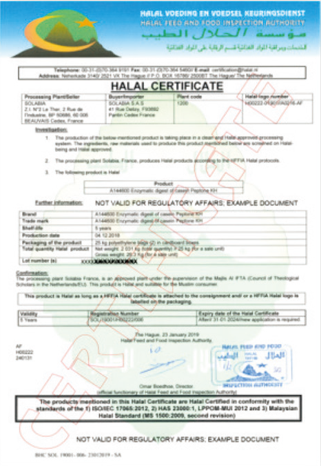
HALAL CERTIFICATE(样本)

▶▶Kosher认证产品一览

厂家编号

产品名称

包装

主要原料

消化酶(来源)

Kosher

Halal

A160100

Papaic digest of soybean meal USP

25 kg

脱脂大豆粉

植物、微生物来源

 经IP(identity-Preserved)处理,达到氨基酸、糖平衡的脱脂大豆粉来源蛋白胨。

A160300

Soy peptone F

25 kg

脱脂大豆粉

植物、微生物来源

 经IP(identity-Preserved)处理,由Solabia的工艺处理和生产的脱脂大豆粉来源蛋白胨。

A160700

Papaic soy peptone

25 kg

脱脂大豆粉

植物、微生物来源

 经IP(identity-Preserved)处理,达到糖、氮、微量元素平衡的发酵用脱脂大豆粉的蛋白胨。

A162100

Soy peptone Crystale

25 kg

脱脂大豆粉

植物、微生物来源

 适合透明高的肉汤培养,含高水平碳水化合物的脱脂大豆来源蛋白胨。

A210100

Wheat peptone

25 kg

小麦

植物、微生物来源

 由小分子肽组成的小麦来源蛋白胨,适合细胞培养。可作为动物产品的替代品,有效培养细菌和酵母。

A220100

Broadbean peptone

25 kg

蚕豆

植物、微生物来源

 对乳酸菌表现出良好的生长作用,在发酵、微生物检测和细胞培养中作为肉类蛋白胨的替代品使用。

A230100

Lupin peptone

25 kg

羽扇豆

植物、微生物来源

 由小分子肽组成,具有应用于发酵、生物制药以及作为动物产品的替代品的细胞培养等各种用途。

A240100

Potato peptone

25 kg

土豆

植物、微生物来源

 具有作为发酵生长培养基、细胞培养补充剂和无动物培养基等各种用途。

A250100

Pea peptone

25 kg

豌豆

植物、微生物来源

 由800 Da或以下小分子组成的蛋白胨。可用作肉类蛋白胨、FBS 和细胞培养补充剂的替代品。

A120200

Yeast extract

25 kg

发芽酵母

植物、微生物来源

 自消化面包酵母(Saccharomyces cerevisiae)的提取物。具有高含量的维生素和氨基酸,被认为是培养基的主要浓缩因子。促进多种微生物

 的生长。

A144100

Enzymatic digest of casein Kosher

25 kg

微生物来源

 其性能设计与Tryptone USP相同,经Kosher认证适用于食品和发酵方面的蛋白胨。

▶▶Kosher/Halal认证产品一览

厂家编号

产品名称

包装

主要原料

消化酶(来源)

Kosher

Halal

A144600

Enzymatic digest of casein KH

25 kg

微生物来源

 与A144100为同一产品,已取得Kosher/Halal认证的高性能酪蛋白胨。促进多种微生物的生长。

A163700

Papaic soy peptone KPH

25 kg

脱脂大豆粉

植物、微生物来源

 与A160700为同一产品,已取得Kosher/Halal认证的脱脂大豆粉来源蛋白胨,。

A253100

Pea peptone KPH

25 kg

豌豆

植物、微生物来源

与A250100为同一产品,已取得Kosher/Halal认证的豌豆来源蛋白胨。

A163300

Soy peptone F KPH

25 kg

脱脂大豆粉

植物、微生物来源

 与A160300为同一产品,已取得Kosher/Halal认证的脱脂大豆粉来源蛋白胨。

A213100

Wheat peptone KPH

25 kg

小麦

植物、微生物来源

与A210100为同一产品,已取得Kosher/Halal认证的小麦来源蛋白胨。

A223100

Broadbean peptone KPH

25 kg

蚕豆

植物、微生物来源

 与A220100为同一产品,已取得Kosher/Halal认证的蚕豆来源蛋白胨。

A233100

Lupin peptone KPH

25 kg

羽扇豆

植物、微生物来源

 与A230100为同一产品,已取得Kosher/Halal认证的羽扇豆来源蛋白胨。

A243100

Potato peptone KPH

25 kg

土豆

植物、微生物来源

 与A240100为同一产品,已取得Kosher/Halal认证的土豆来源蛋白胨。

产品编号 产品名称 产品规格 产品等级

蛋白胨 • 蛋白水解物 应用于微生物培养、发酵、生物制药生产

  • 产品特性
  • 相关资料
  • Q&A
  • 参考文献

应用于微生物培养、发酵、生物制药生产蛋白胨 • 蛋白水解物                              应用于微生物培养、发酵、生物制药生产

蛋白胨 · 蛋白水解物


Solabia Biotechnology是法国Solabia集团下负责研发并生产蛋白胨/蛋白水解物的蛋白胨部门,提供大包装的高质量产品。可作为食品和发酵产品的生产过程中提高微生物检测和疫苗等生物制药生产的细胞培养效率所必需的营养素来使用。生产工厂在法国和巴西均设有多个基地,大幅度降低了缺货的风险,在COVID-19的影响下也能实现持续供应。富士胶片和光可提供40多种经Kosher或Halal认证的,植物、酵母、牛奶酪蛋白、动物来源的蛋白胨产品。

◆特点


● 动物源产品和无动物源产品分开生产

 大量产品已取得Kosher/Halal 认证

 彻底的可溯源性

 常规包装为25 kg的大包装

 设有多个生产基地,降低缺货风险


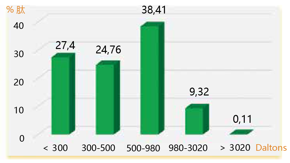
◆牛奶酪蛋白来源产品


厂家编号

产品名称

消化酶

包装

A143100

Tryptone USP NZ

以胰酶为主

25 kg

与大豆蛋白胨或酵母提取物等结合使用,可促进多种微生物的生长。

▶ 分子量分布

蛋白胨 • 蛋白水解物                              应用于微生物培养、发酵、生物制药生产

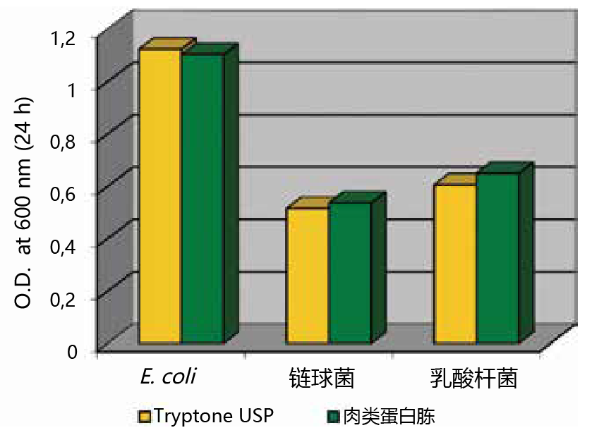
▶ 增殖示例

蛋白胨 • 蛋白水解物                              应用于微生物培养、发酵、生物制药生产

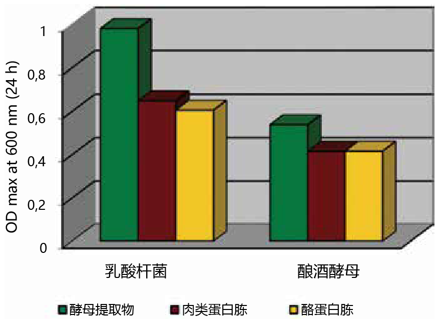
Tryptone USP NZ对各种微生物表现出优异的生长促进作用,可用作通用底物。

培养液:108 cfu/mL

生长培养基:3%蛋白胨+0.25%葡萄糖


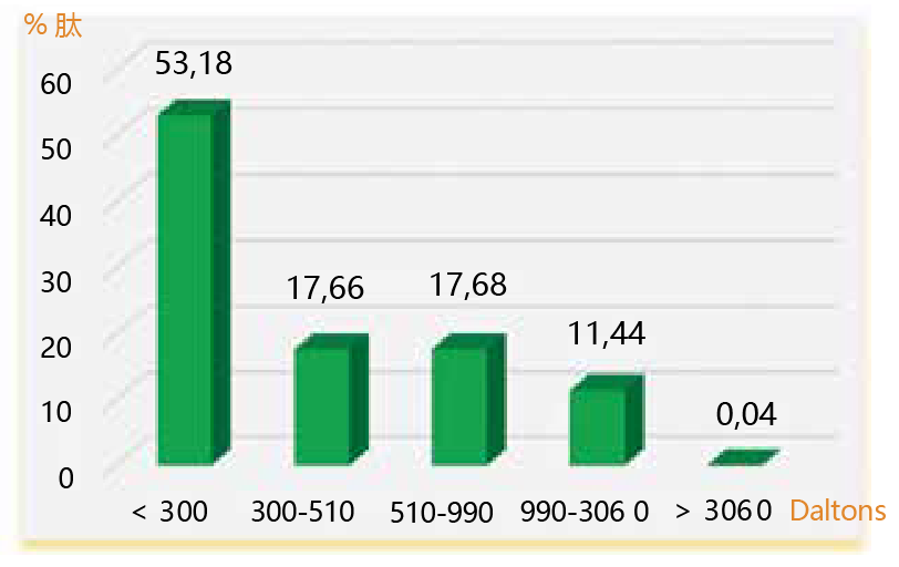
◆酵母来源产品

厂家编号

产品名称

消化酶

包装

A120200

Yeast extract

酿酒酵母

25 kg

与大豆蛋白胨或酵母提取物等结合使用,可促进多种微生物的生长。

▶ 分子量分布


蛋白胨 • 蛋白水解物                              应用于微生物培养、发酵、生物制药生产


▶ 增殖示例


蛋白胨 • 蛋白水解物                              应用于微生物培养、发酵、生物制药生产

酵母提取物(Yeast extract)对含酵母、霉菌等多种微生物表现出良好的生长促进作用。

广泛应用于诊断、发酵、细胞培养等。


培养液:106 cfu/mL

生长培养基:3% 蛋白胨+0.25%葡萄糖


关于Solabia的业务形式

蛋白胨 • 蛋白水解物                              应用于微生物培养、发酵、生物制药生产

※ 富士胶片和光仅销售Diagnostics、Biotechnology的产品。

※ 本页面产品仅供研究用,研究以外不可使用。


产品编号 产品名称 产品规格 产品等级

蛋白胨•蛋白水解物 应用于微生物培养、发酵、生物制药生产

  • 产品特性
  • 相关资料
  • Q&A
  • 参考文献

应用于微生物培养、发酵、生物制药生产蛋白胨•蛋白水解物                              应用于微生物培养、发酵、生物制药生产

蛋白胨·蛋白水解物

Solabia Biotechnology是法国Solabia集团下负责研发并生产蛋白胨/蛋白水解物的蛋白胨部门,提供大包装的高质量产品。可作为食品和发酵产品的生产过程中提高微生物检测和疫苗等生物制药生产的细胞培养效率所必需的营养素来使用。富士胶片和光可提供40多种经Kosher或Halal认证的,植物、酵母、牛奶酪蛋白、动物来源的蛋白胨产品。

◆特点


● 动物源产品和无动物源产品分开生产

 大量产品已取得Kosher/Halal 认证

 彻底的可溯源性


蛋白胨•蛋白水解物                              应用于微生物培养、发酵、生物制药生产

◆产品列表(豌豆、羽扇豆来源)

厂家编号

产品名称

包装

消化酶

无动源成分

无过敏原

Kosher

Hala

A250100

Pea peptone

25 kg

植物来源
微生物来源

×

  由800 Da或以下小分子量组成的豌豆来源蛋白胨。肉类蛋白胨、FBS和细胞培养补充剂的替代品。

A253100

Pea peptone KPH

25 kg

植物来源
微生物来源

  与A250100为同一产品,已取得Kosher・Halal认证的豌豆来源蛋白胨。

A220100

Broardbean peptone

25 kg

植物来源
微生物来源

×

  对乳酸菌可呈现优异增殖的蚕豆来源蛋白胨。在发酵、微生物检测、细胞培养中,作为肉类来源蛋白胨的替代品发挥功能。

A223100

Broardbean peptone KPH

25 kg

植物来源
微生物来源

  与A220100为同一产品,已取得Kosher/Halal认证的羽扇豆来源蛋白胨。

※ 本页面产品仅供研究用,研究以外不可使用。


产品编号 产品名称 产品规格 产品等级

泪液粘蛋白检测试剂盒 Tear Mucin Assay kit

  • 产品特性
  • 相关资料
  • Q&A
  • 参考文献

泪液粘蛋白检测试剂盒泪液粘蛋白检测试剂盒                              Tear Mucin Assay kit

Tear Mucin Assay kit

  本试剂盒的原理是在碱性条件下,O-聚糖通过β氢脱离反应从核心蛋白中脱离的同时,在糖链还原末端标记荧光,根据荧光强度检测泪液中的粘蛋白含量。


◆背景

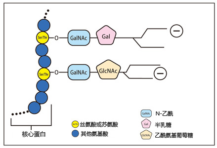
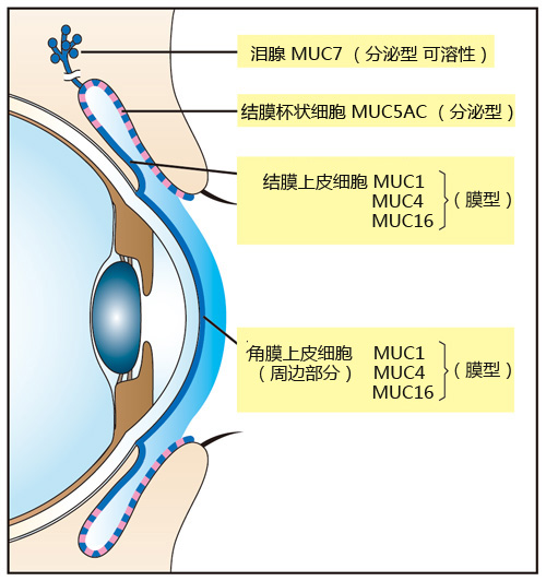
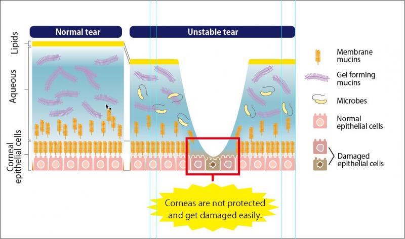
  粘蛋白是泪液和眼表上皮顶端细胞膜的主要组分,由O-糖链结合核心蛋白而成。其中,核心蛋白是由10-80氨基酸残基,通过高度O-糖基化的丝氨酸和苏氨酸残基串联重复结构域而组成。,而具有亲水性质的O-聚糖占粘蛋白重量的一半以上(图1)。重糖基化的粘蛋白具有高负电荷和亲水性,该特性具有阻止病原体粘附或穿透进入上皮的功能(图2)。干眼病患者眼表面粘蛋白及其分泌均发生改变(图2)。在眼表上皮,存在三种类型的粘蛋白,由结膜杯状细胞表达的大凝胶形成的粘蛋白MUC5AC,一些来自泪腺腺泡的细胞表达小溶性粘蛋白MUC7,角膜和结膜上皮细胞表达膜相关粘蛋白MUC1,4和16(图2)。

泪液粘蛋白检测试剂盒                              Tear Mucin Assay kit


 图1 粘蛋白的构造 


泪液粘蛋白检测试剂盒                              Tear Mucin Assay kit


2 围绕在角膜表面的粘蛋白表达以及泪液粘蛋白的屏障功能


泪液粘蛋白检测试剂盒                              Tear Mucin Assay kit


图3 泪液粘蛋白的防护机能

  泪液中粘蛋白的减少被认为是干眼症的病因。泪液中的粘蛋白可大致分为两大类,分别是分泌型粘蛋白以及膜型粘蛋白(图2)。分泌型粘蛋白包括MUC5AC(由结膜杯状细胞分泌)、MUC7(由泪腺分泌);膜型粘蛋白包括MUC1、MUC4、MUC16(存在于角膜上皮的细胞膜上)等。


◆特点


●  本试剂盒的原理是在碱性条件下,O-聚糖通过β氢脱离反应从核心蛋白中脱离的同时,在糖链还原末端标记荧光,根据荧光强度检测

  泪液中的粘蛋白含量。

  使用检测泪液量的泪液分泌试纸可检测泪液中粘蛋白的含量。
  可用于针对干眼症等眼部问题的药物及保健食品的研发以及眼科相关研究。


◆试剂盒组成


NO.

组成

规格

数量

贮存温度

1

提取液

 30 mL 


4~10℃

2

凝胶过滤载体

 45 mL

3

标准液

(100 µg/mL 

N-乙酰)

1.0 mL 

4

试剂 A

0.3 mL

5

试剂 B

1.5 mL

6

反应停止液

15 mL

7

空柱

1 mL用

50

8

离心管

50

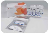
该试剂盒包含检测数目泪液黏蛋白所有组件


◆应用


  用于测定泪液中粘蛋白含量。

测定原理


  粘蛋白属于高分子(1000 kda-10000 kda)重糖基化蛋白家族,主要由富含苏氨酸与丝氨酸的核心蛋白组成。糖链N-乙酰半乳糖胺(GalNAc)的还原末端与翻译后的O-糖基化氨基酸连接(图1)。在碱性条件下,O-聚糖通过β氢脱离反应从核心蛋白中脱离的同时,在高温下糖链还原末端与2-氰基乙酰胺(2-CAN)反应产生强烈荧光缩合物,根据荧光强度检测泪液中的粘蛋白含量。

检测步骤


● 本产品专为荧光(96孔板)测量而设计。

● 使用荧光读板机可以测量100个样品。

● 使用分光光度计测量时,请使用微细胞。


样品制备

● 使用Schirmer氏泪液分泌条收集泪液5分钟。

● 储存泪液分泌条收集的泪液时,请放入微量试管中,并在4℃保存,同时避免冻结。


泪液粘蛋白的测定

1、将泪液分泌条转移至微量试管中,加入 200 μL 缓冲液洗脱,并在室温下提取粘蛋白1小时。

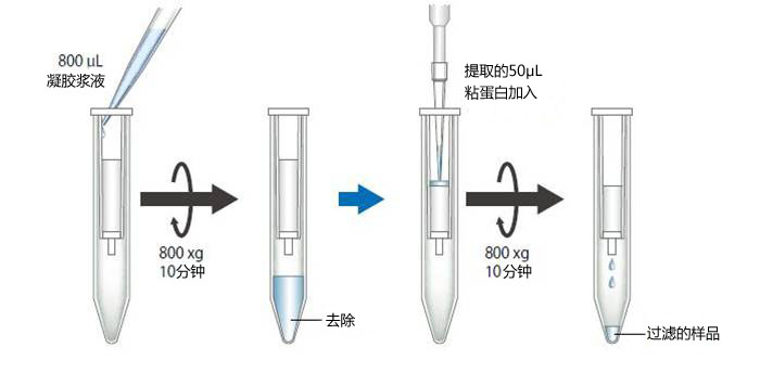
2、在步骤1的1小时内,准备空柱,并将空柱放入离心管中。搅拌凝胶浆液至均匀后,将 800 μL 凝胶浆液加入到空柱中,并在室温下以 800×g 

   离心 10 分钟,(推荐使用具有摆动转子系统的离心机。)去除管中底部液体。

3、将步骤1用泪液分泌条提取的 50 μL 粘蛋白加入到步骤2凝胶的上部。

4、在室温下以 800×g 离心加有样品的柱子 10 分钟,并收集过滤的样品组分(约50 μL)。


泪液粘蛋白检测试剂盒                              Tear Mucin Assay kit


5、将每次通过的组分(4)或标准溶液(①~⑧)的 20 μL 转移到另一个微量试管(500 μL)中。在试管中加入 24 μL 混合试剂(使用前,

   将试剂A和试剂B以 1:5 的体积混合)。混合后,将试管加热至100℃ 30分钟。

6、将试管冷却至室温后,加入 200 μL 终止溶液,并用涡旋混合器混匀。

7、将100 μL溶液放入96孔黑色孔板中,设置波长(荧光:336 nm,发射:383 nm)后用荧光读板机测量。

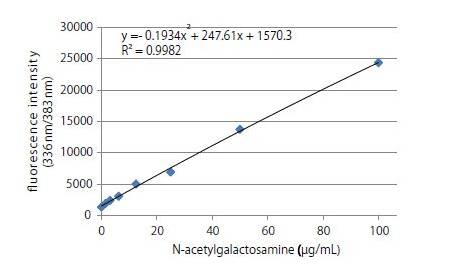
8、按照如下稀释浓度创建标准曲线。


  构建校准曲线,并从标准曲线中读取样品浓度。


标准曲线:


泪液粘蛋白检测试剂盒                              Tear Mucin Assay kit


参考数据:

建议测量 Ex 时使用 336 nm 波长,测量 Em 时使用 383 nm,但使用具有干涉滤光片系统的荧光板作为前导时,可能无法测量荧光波长,这种情况下,需将荧光波长调到较长数值。


相关产品信息请点击:粘蛋白检测试剂盒


参考文献

[1]


Gipson IK, Hori Y, Argüeso P. Character of ocular surface mucins and their alteration in dry eye disease.(2004)Ocul Surf., Apr;2(2):131-48


[2]


Argüeso P, Balaram M, Spurr-Michaud S, Keutmann HT, Dana MR, Gipson IKGipson IK et al., Decreased levels of the goblet cell mucin MUC5AC in tears of patients with Sjögren syndrome. (2002) Invest Ophthalmol Vis Sci, April; 43(4): 1004-11


[3]


Uchino Y, Uchino M, Yokoi N, Dogru M, Kawashima M, Okada N, Inaba T, Tamaki S, Komuro A, Sonomura Y, Kato H, Argüeso P, Kinoshita S, Tsubota, Alteration of tear mucin 5AC in office workers using visual display terminals: The Osaka Study. (2014) JAMA Ophthalmol., Aug;132(8):985-92


[4]


natomi T, Spurr-Michaud S, Tisdale AS, Zhan Q, Feldman ST, Gipson IK. Expression of secretory mucin genes by human conjunctival epithelia. (1996) Invest Ophthalmol Vis Sci., Aug;132(8):985-92


[5]


Susumu Honda, Yoshikazu Matsuda, Masaye Takahashi, and Kazuaki Kakehi Fluorimetric Determination of Reducing Carbohydrates with2-Cyanoacetamide and Application to Automated Analysis of Carbohydrates as Borate Complexes. (1980) Analytica Chimica, Vol. 52, No. 7


[6]


Crowther RS, Wetmore RF: Fluorometric assay of O-linked glycoproteins by reaction with 2-cyanoacetamide. Anal Biochem 163: 170-174, 1987.


[7]


Okazaki Y, Han Y, Kayahara M, Watanabe T, Arishige H, Kato N. Consumption of curcumin elevates fecal immunoglobulin A, an index of intestinal immune function, in rats fed a high-fat diet. J Nutr Sci Vitaminol (2010); 56(1): 68-71.


[8]


Yukako Okazaki, Hiroyuki Tomotake, Kazuhisa Tsujimoto, Masahiro Sasaki,and Norihisa Kato. Consumption of a Resistant Protein, Sericin, Elevates Fecal Immunoglobulin A, Mucins, and Cecal Organic Acids in Rats Fed a High-Fat Diet. (2011) The Journal of Nutrition , 21, 10.3945/ jn.111.144246.


[9]


Zaki Utama & Yukako Okazaki & Hiroyuki Tomotake & Norihisa Kato, Tempe Consumption Modulates Fecal Secondary Bile Acids, Mucins, Immunoglobulin A, Enzyme Activities, and Cecal Microflora and Organic Acids in Rats. Plant Foods Hum Nutr (2013) 68: 177–183


产品编号 产品名称 产品规格 产品等级
MUC01  Tear Mucin Assay Kit (O-Glycan Assay Method 1 KIT [50TEST] 

粘蛋白检测试剂盒 Mucin Assay Kit

  • 产品特性
  • 相关资料
  • Q&A
  • 参考文献

粘蛋白检测试剂盒粘蛋白检测试剂盒                              Mucin Assay Kit

Mucin Assay Kit

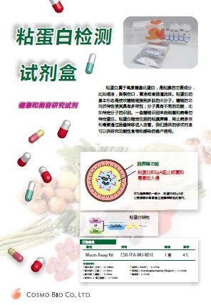
——健康和美容研究试剂


背景


粘蛋白属于高度糖基化蛋白质家族,是粘膜的主要成分,例如唾液,泪液,胃液,肠液。粘蛋白的 基本构型是枝状糖链连接到多肽的大分子。糖链的非均质特性使其具有多样性,分子具有各种功能,如特异性分子识别。
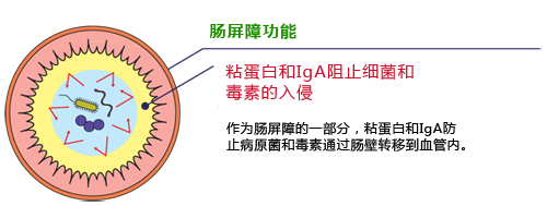
  一些糖链识别病毒和细菌的特定蛋白质。粘蛋白锚定在肠中粘膜屏障功能中,阻止病原体和毒素通过肠壁进入血管中。该试剂盒可用于检测粪便中的粘蛋白含量。
  粘蛋白属于高分子(1000 kda-10000 kda)高度糖基化蛋白的家族。蛋白质核内的粘蛋白结构域富含苏氨酸、丝氨酸和羟脯氨酸,糖链的还原末端(GalNAc)通过翻译后O-糖基化与这些氨基酸频繁连接。该试剂盒具有确定粪便
粘蛋白含量组分的功能。

  粪便粘蛋白测定可用作肠屏障功能的指标。该试剂盒利用荧光检测来测定粘蛋白的含量。

  我们提供的该试剂盒可以供研究功能性食物和感染的客户使用。

粘蛋白检测试剂盒                              Mucin Assay Kit

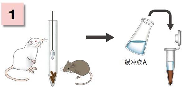
步骤1:从粪便中提取和部分纯化粘蛋白。

步骤2:粘蛋白O-糖苷连接的寡糖链的测定通过稀碱进行β-消除,形成糖链的还原端。还原碳水化合物在高温下进行荧光标记以产生荧光缩合

            物。


粘蛋白的结构

粘蛋白检测试剂盒                              Mucin Assay Kit

粘蛋白检测试剂盒                              Mucin Assay Kit

 试剂盒组分


组件

规格

数量

储存条件

缓冲溶液A

100 mL

3

4-10℃

缓冲溶液B

25 mL

1

缓冲溶液C

25 mL

1

试剂A

1.0 mL

1

试剂B

1.5 mL

2

标准溶液

(GalNA,250 μg/mL)

1.0 mL

1

酶溶液

1.5 mL

1

◆优点特色


●  粘蛋白和 IgA 能阻止细菌和毒素的入侵

●  作为肠屏障的一部分,粘蛋白和 IgA 能防止病原菌和毒素通过肠壁转移到血管内。


测定理论
  通过对粪便中的 β-葡萄糖苷酶热变性防止粪便粘蛋白降解
  ↓
  粗提取粘蛋白
  ↓
  用酶溶液分解淀粉
  ↓
  用乙醇沉淀纯化粘蛋白
  ↓
  将试剂A加入纯化的粘蛋白中,并在碱性条件下进行热处理。 (试剂A将与粘蛋白的糖还原末端反应,并在碱处理后发出荧光)
  ↓
  加入缓冲液C并测定荧光强度(激发336nm,荧光383nm)

  (具体操作请看相关资料)

◆案例应用


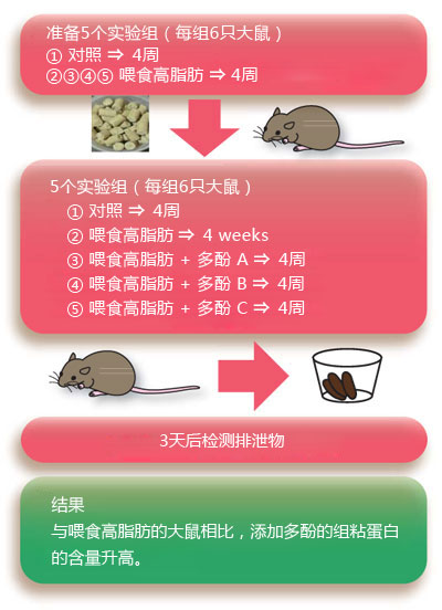
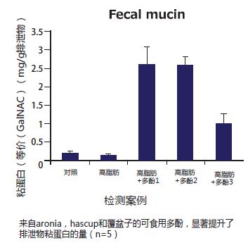
喂食高脂肪食物的大鼠肠屏障部分注射多酚的效果

粘蛋白检测试剂盒                              Mucin Assay Kit

粘蛋白检测试剂盒                              Mucin Assay Kit

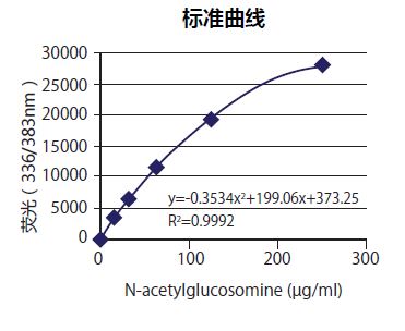
图1 粘蛋白定量标准曲线

粘蛋白检测试剂盒                              Mucin Assay Kit

图2 粪便中的粘蛋白含量

应用

  用于测定粪便中的粘蛋白含量。


相关资料

粘蛋白检测试剂盒                              Mucin Assay Kit

Mucin_Assay_Kit.

◆操作步骤


● 本产品是专为荧光酶标仪测量设计(96孔板)。
● 当使用荧光读板机时,可测量100个样品。
● 当使用分光光度计测量时,请使用微细胞。


缓冲液A的制备
用100 mL蒸馏水溶解1块片剂。
粪便粉末的制备:粪便冻干并在研钵中研磨,储存在-20℃备用。

II.粪便粘蛋白的测定
  1称量100 mg粪便粉末放入微量试管(2 mL)中,加入1 mL缓冲液A,然后用涡流混合器混合30秒钟。


粘蛋白检测试剂盒                              Mucin Assay Kit


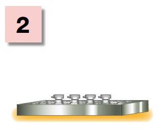
  2在95℃下加热试管10分钟使细菌的糖苷酶变性。


粘蛋白检测试剂盒                              Mucin Assay Kit


  3在37℃下加热90分钟,从粪便中提取粘蛋白。

  4在4℃下离心20,000×g 15分钟。
  5将200 μL上清液转移到另一个微量试管中,加入200 μL缓冲液B,用涡流混合器混合溶液。

  6加入10 μL酶溶液混合,在50℃下加热试管20分钟分解淀粉。
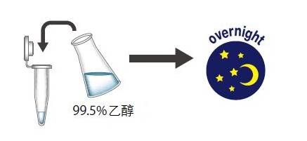
  7将离心管冷却至室温,加入125 μL的99.5%乙醇,混合后,在-20℃下静置过夜。


粘蛋白检测试剂盒                              Mucin Assay Kit


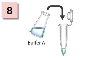
  8次日,在4℃下20,000×g离心10分钟,去上清。向沉淀加入1mL缓冲液A,重溶沉淀。(样品溶液)


粘蛋白检测试剂盒                              Mucin Assay Kit


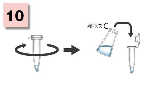
  9、将20 μL样品溶液和标准溶液转移到另一个微量试管(500 μL)中,加入24 μL试剂混合物(使用前以1:5混合试剂A和试剂B),混合后, 100℃下加热微量试管30分钟。

  10、冷却至室温,加入200 μL缓冲液C,用涡流混合器混合。


粘蛋白检测试剂盒                              Mucin Assay Kit


  11将100 μL溶液转移到96孔黑色孔板中,并在波长(例如:336 nm em:383 nm)实用荧光读板机测量荧光强度。
  12通过如下所示的连续稀释创建标准曲线。 通过这些点绘制平滑曲线以构建标准曲线。 通过标准曲线读取样品的浓度。
  13计算1 g粪便中的粘蛋白含量。步骤(12)测得的值的50倍是1g粪中的粘蛋白含量。

Calibrator Number

Quantaity of Standard

Quantaity of Dilution Buffer 

Final Concentration

1

500 μL of Standard Solution

0

250 μg/mL

2

500 μL of Calibrator 1

500 μL

125 μg/mL

3

500 μL of Calibrator 2

500 μL

63 μg/mL

4

500 μL of Calibrator 3

500 μL

31.5 μg/mL

5

500 μL of Calibrator 4

500 μL

15.75 μg/mL

6

500 μL of Calibrator 5

500 μL

7.875 μg/mL

7

0

500 μL

0

工作校准品:N-乙酰葡萄糖胺250 μg/ mL
● 通过连续稀释创建标准曲线,如下图所示。
● 剩余的未稀释校准品应储存在2-10°C。
● 稀释校准品稳定,可在2-10°C保存1个月。

参考文献


[1]

Susumu Honda,Yoshikazu Matsuda, Masaye Takahashi, and Kazuaki KakehiFluorimetric Determination of Reducing Carbohydrates with2-Cyanoacetamide and Application to Automated Analysis of Carbohydrates as Borate Complexes.(1980) Analytical Chemistry, Vol.52, No. 7


[2

Bovee-OudenhovenIM, Termont DS, Heidt PJ, et al.: Increasing the intestinal resistance of rats to the invasive pathogen Salmonella enteritidis: additive effects of dietary lactulose and calcium. Gut 40: 497-504, 1997.


[3

Crowther RS, Wetmore RF: Fluorometric assay of O-linked glycoproteins by reaction with 2-cyanoacetamide. Anal Biochem 163: 170-174, 1987.


[4

Okazaki Y, Han Y, Kayahara M, Watanabe T, Arishige H, Kato N. Consumption of curcumin elevates fecal immunoglobulin A, an index of intestinal immune function, in rats fed a high-fat diet. J NutrSciVitaminol (2010);56(1):68-71.

[5

YukakoOkazaki,HiroyukiTomotake, Kazuhisa Tsujimoto, Masahiro Sasaki,andNorihisa Kato. Consumption of a Resistant Protein, Sericin, Elevates Fecal Immunoglobulin A, Mucins, and Cecal Organic Acids in Rats Fed a High-Fat Diet. (2011) The Journal of Nutrition, 21, 10.3945/jn.111.144246.

[6

ZakiUtama,Yukako Okazaki, Hiroyuki Tomotake, Norihisa Kato, Tempe Consumption Modulates Fecal Secondary Bile Acids, Mucins, Immunoglobulin A, Enzyme Activities, and CecalMicroflora and Organic Acids in Rats. Foods Hum Nutr(2013) 68:177-183

产品编号 产品名称 产品规格 产品等级
CSR-FFA-Mu-K01E Mucin Assay Kit
粘蛋白检测试剂盒
1盒

蛋白胨

  • 产品特性
  • 相关资料
  • Q&A
  • 参考文献

蛋白胨蛋白胨

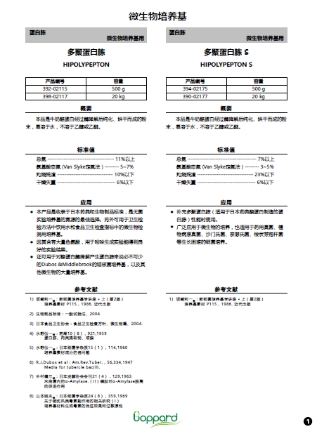
多聚蛋白胨

HIPOLYPEPTON

◆概要


  本品是牛奶酪蛋白经过酶降解后纯化、烘干而成的粉末,易溶于水,不溶于乙醇或乙醚。

◆标准值

总氮

11%以上

氨基酸态氮(Van Slyke定氮法)

5~7%

灼烧残渣

10%以下

干燥失重

6%以下

◆应用


1) 本产品是收录于日本药典和生物制品标准,是无菌实验培养基的氮源的理想选择。另外可用于卫生检验方法中饮用水和食品卫生检查指标中

       微生物检测用培养基

2) 因其含有大量色氨酸,用于吲哚生成实验能得到良好的实验结果。

3) 还可用于对酪蛋白酶降解产生蛋白胨来说必不可少的Dubos &Middlebrook的结核菌培养基,以及其他微生物的大量培养基。

—————————————————————————————————————————————————————————————————————

多聚蛋白胨 S

HIPOLYPEPTON S

◆概要


  本产品是脱脂大豆经过酶降解后纯化、烘干而成的植物性蛋白胨粉末,易溶于水,不溶于乙醇或乙醚。

◆标准值

总氮

7%以上

氨基酸态氮(Van Slyke定氮法)

3~5%

灼烧残渣

23%以下

干燥失重

6%以下

◆应用

1) 补充多聚蛋白胨(适用于日本药典酪蛋白制造的蛋白胨)性能时使用;

2) 广泛应用于微生物的培养,也适用于药用真菌、植物病原真菌、沙门氏菌、奈瑟氏菌、梭状芽孢杆菌等生长困难的细菌培养。

—————————————————————————————————————————————————————————————————————


多聚蛋白胨 N

HIPOLYPEPTON N

◆概要


  本产品是大豆提纯产品经过微生物来源的酶降解后纯化、烘干而成的植物性蛋白胨粉末,其性能与多聚蛋白胨相同。

◆标准值


总氮

12%以上

氨基酸态氮(Van Slyke定氮法)

5~7%

灼烧残渣

15%以下

干燥失重

6%以下

◆应用

非动物性蛋白胨,用途和多聚蛋白胨相同。

—————————————————————————————————————————————————————————————————————

多聚蛋白胨 NS

HIPOLYPEPTON NS

◆概要


  本产品是使用微生物酶降解大豆提纯产品后纯化、烘干而成的植物性蛋白胨粉末。性能和多聚蛋白胨S相同。

◆标准值

总氮

7%以上

氨基酸态氮(Van Slyke定氮法)

3~5%

灼烧残渣

23%以下

干燥失重

9%以下

◆应用

非动物性蛋白胨的用途和多聚蛋白胨S相同。

—————————————————————————————————————————————————————————————————————

高酪蛋白酸“DAIGO”

HICASAMONO ACIDS DAIGO

◆概要

  本产品是牛奶酪蛋白的酸降解物。其中,蛋白质几乎均被分解成氨基酸,而且几乎不含蛋白质以外的营养源和生长因子。它可用作细菌生理学、遗传学等基础领域里使用的培养基的纯氮源,或作为定量维生素(特别是B12)培养基里的氮源。还可以像MH琼脂培养基等不适合酶消化蛋白胨使用的培养基作为营养源来使用。

◆标准值

总氮

7%以上

氨基酸态氮(Van Slyke定氮法)

54~83%

灼烧残渣

52%以下

干燥失重

8%以下

氯化物(NaCl)

28~40%

pH值

6~7


蛋白胨

蛋白胨和酵母粉

多聚蛋白胨

HIPOLYPEPTON

参考文献


1) 坂崎利一等:新细菌培养基学讲座 · 上(第2版), 培养基素材 P115,1986. 近代出版

2) 生物制品标准:一般试验法、2004

3) 日本食品卫生协会:食品卫生检查方针、微生物篇、2004.

4) 水野伝一等:药局10(8),921,1959   

      蛋白胨、肉类提取物、琼脂

5) 水野伝一等:日本细菌学杂质15(1),114,1960

      培养基素材成分的各问题

6) R.J.Dubos et al:Am.Rev.Tuber.,56,334,1947

      Media for tubercle bacilli.

7) 外村健三等:日本发酵协会会刊21(4),129,1963

      米曲菌内的α-Amylase.(II)磷肽对α-Amylase脱离的促进作用

8) 山本昭夫等:日本细菌学杂质24(8),359,1969

      关于破伤风类毒素副作用的相关研究(I)

      培养基材料生成毒素的促进效果和过敏原性

多聚蛋白胨 S

HIPOLYPEPTON S

参考文献


1) 坂崎利一他:新细菌培养基学讲座 · 上(第2版),  培养基素材 P115,1986. 近代出版

高酪蛋白酸“DAIGO”

HICASAMONO ACIDS DAIGO

参考文献


1) 坂崎利一等:新细菌培养基学讲座 · 上(第2版),  培养基素材 P115,1986. 近代出版

产品编号 产品名称 产品规格 产品等级
392-02115 多聚蛋白胨 500g 500 g
398-02117 多聚蛋白胨 20kg 20 kg
394-02175 多聚蛋白胨 S 500g 500 g
390-02177 多聚蛋白胨 S 20kg 20 kg
397-02121 多聚蛋白胨 N 300g 300 g
395-02127 多聚蛋白胨 N 10kg 10 kg
393-02101 多聚蛋白胨 NS 300g 300 g
391-02107 多聚蛋白胨 NS 10kg 10 kg
393-02145 高酪蛋白酸“DAIGO” 500g 500 g
399-02147 高酪蛋白酸“DAIGO” 10kg 10 kg

pCAG-Fc(Fc融合蛋白表达载体) pCAG-Hyg hIgG1-Fc

  • 产品特性
  • 相关资料
  • Q&A
  • 参考文献

重组抗体和嵌合蛋白研究的理想选择pCAG-Fc(Fc融合蛋白表达载体)                              pCAG-Hyg hIgG1-Fc

pCAG-Fc (Fc 融合蛋白表达载体)

  pCAG 是动物细胞中基因表达载体。通过 CAG 启动子的运作,在动物细胞内的目标蛋白可高表达。

  pCAG-Hyg/Neo IgG-Fc 是在具有各种铰链序列的 IgG-Fc 区中,C末端带有多克隆位点(MCS)的载体,可使附加在 IgG-Fc 区中的目的重组蛋白表达出来。由于 MCS 的N末端已插入分泌信号序列,Fc 融合嵌合蛋白可分泌在培养上清中。

  Fc 融合嵌合蛋白在保留原有的蛋白活性的同时,具有半衰期长、在哺乳类细胞中的高表达、可简便提纯 Protein A和 Protein G等特点,主要应用于蛋白质的功能解析和重组蛋白的基础研究中。


◆特点


● CAG 启动子诱导高表达

● 优化耐药基因的表达水平

● 已插入分泌信号序列

● 多种限制性酶适用


载体图谱


pCAG-Fc(Fc融合蛋白表达载体)                              pCAG-Hyg hIgG1-Fc

※1 可能因蛋白质的性质,而不具分泌性。※2 本品仅供实验研究用。


可用的限制性酶列表


Vector

Acc I

BamH I

Cla I

EcoR I

EcoR V

Hind Ⅲ

Pst I

Sal I

Sma I

Spe I

Xho I

pCAG-Hyg hIgG1-Fc

pCAG-Hyg mIgG1-Fc

pCAG-Hyg mIgG2a-Fc

pCAG-Hyg mIgG2b-Fc

pCAG-Hyg rIgG2a-Fc

pCAG-Hyg rIgG2b-Fc

pCAG-Neo hIgG1-Fc

pCAG-Neo mIgG1-Fc

pCAG-Neo mIgG2a-Fc

pCAG-Neo mIgG2b-Fc

pCAG-Neo rIgG2a-Fc

pCAG-Neo rIgG2b-Fc

大部分限制性酶可用

新标准抗体纯化树脂 性价比高!!


KANEKA KanCapA™ 为 Protein A 亲和层析树脂。用于从活体样本及细胞培养上清纯化免疫球蛋白及包含Fc段的重组抗体。

本品由高交联纤维素微珠及新研发的耐碱 Protein A 配体构成,可用于 IgG 的亲和纯化。

pCAG-Fc(Fc融合蛋白表达载体)                              pCAG-Hyg hIgG1-Fc

产品编号 产品名称 产品规格 产品等级
165-27741 pCAG-Hyg hIgG1-Fc 20μg
162-27751 pCAG-Hyg mIgG1-Fc 20μg
169-27761 pCAG-Hyg mIgG2a-Fc 20μg
166-27771 pCAG-Hyg mIgG2b-Fc 20μg
163-27781 pCAG-Hyg rIgG2a-Fc 20μg
160-27791 pCAG-Hyg rIgG2b-Fc 20μg
163-27801 pCAG-Neo hIgG1-Fc 20μg
160-27811 pCAG-Neo mIgG1-Fc 20μg
167-27821 pCAG-Neo mIgG2a-Fc 20μg
164-27831 pCAG-Neo mIgG2b-Fc 20μg
161-27841 pCAG-Neo rIgG2a-Fc 20μg
168-27851 pCAG-Neo rIgG2b-Fc 20μg
114-01071 KANEKA KanCapATM 2mL(net 1mL)
110-01073 KANEKA KanCapATM 10mL(net 5mL)
118-01074 KANEKA KanCapATM 50mL(net 25mL)

PROTEOSTAT® 蛋白聚集检测 PROTEOSTAT® Protein aggregation assay

  • 产品特性
  • 相关资料
  • Q&A
  • 参考文献

PROTEOSTAT® 蛋白聚集检测                              PROTEOSTAT® Protein aggregation assayPROTEOSTAT® 蛋白聚集检测

  某些肽链在经过错误折叠后很容易形成聚集,在生物技术和制药领域,这些聚集会严重影响蛋白的生产和应用。许多以蛋白多肽为基础研发的药物具有极大的应用前景,可是蛋白因错误折叠而导致构象发生变化形成聚集,使蛋白失活,最终影响药物疗效,造成不可估计的损失。污染物导致的蛋白聚集包括宿主细胞蛋白、非蛋白微粒和蛋白的损伤形式。

  基于此 Enzo Life Sciences 推出了新产品 PROTEOSTAT® Protein Aggregation Assay,内含红色荧光信号染料,可特异性结合聚集蛋白,进行检测。具有广泛的检测范围,可有效检测可见和不可见的蛋白微粒。


 ◆原理

  蛋白聚集严重影响以蛋白为基础研发的药物。在药物制剂中,蛋白聚集在生物活性和免疫原性方面影响药效。

  蛋白聚集发生在生产过程的各个阶段包括细胞培养、纯化、生产、储存、运输等方面。制药工业希望在生物工艺中找到新的方法,可用于检测、追踪、定量分析影响蛋白聚集的因素。

  近年来以冻干稳定形式存在的蛋白聚集体作为标准品,可以准确地定量检测蛋白聚集,加上新颖的只需在酶标仪里即可实验的 PROTEOSTAT® protein aggregation assay,可对蛋白检测方法进行优化。

  在这篇文章中,使用已知浓度的聚集 IgG 标准品作为标准曲线,通过 PROTEOSTAT® protein aggregation assay/standards 对 IgG 的聚集水平进行检测,集中分析了制药工业中常见的3种聚集形式。

  ●    热诱导聚集

  ●    机械诱导聚集

  ●    冷冻和反复冻融诱导聚集


PROTEOSTAT® 蛋白聚集检测                              PROTEOSTAT® Protein aggregation assay

◆优点特色


 ● 高通量筛选方法用于流式细胞仪平台监测蛋白稳定性

 ● 在荧光微孔板中即可进行简单、灵敏、均一地检测

 ● 不需分离目的蛋白或稀释样品

 ● 与传统蛋白聚集检测染料相比更灵敏,信号更明亮

 ● 优化的缓冲液和辅料用于蛋白制剂研发

 ● 检测灵敏度可低至亚微摩尔级别,含量在1%-5%的聚集蛋白也能被检测出

 ● 在广泛的 PH(4-10)和离子强度范围都可使用,提供至少两个数量级的线性动态范围

 ● 使用 PROTEOSTAT® 蛋白聚集检测标准品可对溶液中的聚集蛋白精确定量

 ● 可检测蛋白多肽聚集程度,可用于筛选蛋白聚集激活剂或抑制剂


在蛋白聚集实验研究中,将 PROTEOSTAT® protein aggregation assay 和 Synergy Mx Multi-Mode Microplate reader 联用,可适合用于监测由温度、机械损伤和反复冻融所引起的蛋白聚集。

稳定的蛋白颗粒作为标准品可快速建立标准曲线,加快定量分析蛋白聚集响应。这个检测方法能够可靠地检测到浓缩抗体制剂中小于 0.2% 的聚集蛋白,线性动态范围为2个数量级。

PROTEOSTAT® protein aggregation assay 和 PROTEOSTAT® protein aggregation standards 检测蛋白聚集特点在于操作简单,能够检测低水平的蛋白聚集情况,只需 30 分钟,就能够得到完整的分析结果。


案例一


PROTEOSTAT® 蛋白聚集检测                              PROTEOSTAT® Protein aggregation assay


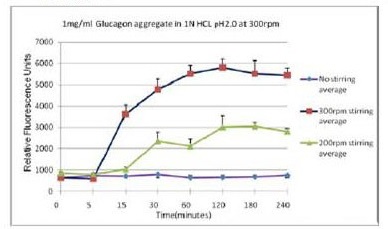
不同搅拌速率对蛋白聚集的影响


案例二


PROTEOSTAT® 蛋白聚集检测                              PROTEOSTAT® Protein aggregation assay


蛋氨酸被氧化后可抑制蛋白聚集

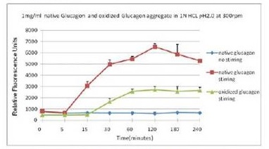
使用本产品检测后,可知搅拌速度越快,蛋白聚集程度越大。

蓝色曲线为对照,没有搅拌速度在 300 rpm 下,绿色曲线与红色曲线分别表示蛋氨酸被氧化后的蛋白聚集情况与天然蛋白的聚集情况。使用本产品检测后,可知蛋氨酸被氧化后可抑制蛋白聚集。

使用流式细胞仪结合PROTEOSTAT® 蛋白聚集分析试剂盒使用例


PROTEOSTAT® 蛋白聚集检测                              PROTEOSTAT® Protein aggregation assay PROTEOSTAT® 蛋白聚集检测                              PROTEOSTAT® Protein aggregation assay PROTEOSTAT® 蛋白聚集检测                              PROTEOSTAT® Protein aggregation assay


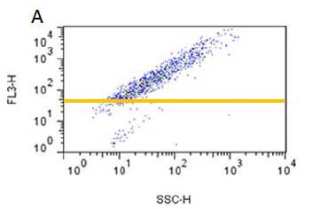
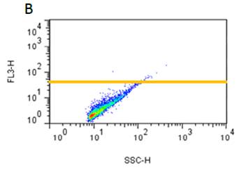
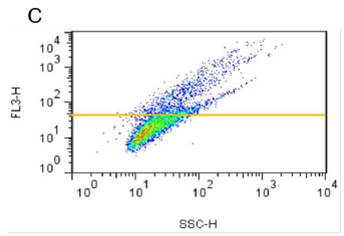
A PROTEOSTAT® 蛋白聚集分析试剂盒检测 IgG 聚集标准品 B PROTEOSTAT® 蛋白聚集分析试剂盒检测硅油滴 C PROTEOSTAT® 蛋白聚集分析试剂盒检测 IgG 聚集标准品和硅油滴的混合物。

使用流式细胞仪结合 PROTEOSTAT® 蛋白聚集分析试剂盒检测 IgG 聚集标准品,可与其他非蛋白微粒如油滴区别开来,且无须考虑样品微粒大小(经检测 >97% 的 IgG 聚集标准品都小于2μm)。

案例・应用

材料和方法


Enzo Life Sciences 推出了 PROTEOSTAT® protein aggregation assay 和 PROTEOSTAT® protein aggregation standards,利用荧光分子信号染料可特异性结合聚集蛋白和多肽,在微孔板中即可检测。

探针在溶液中是不发出荧光的,当与聚集蛋白的四级结构结合时会发出明亮的荧光。一旦聚集标准品发生重组,它们将含有0-12.5% 的聚集蛋白,总蛋白浓度维持在1mg/mL。

通过粒子分析成像鉴定标准品发现90%的蛋白聚集体长度为2-10 μm,约9%的蛋白聚集体长度为 10-20 μm,约 0.7% 的蛋白聚集体为长度大于 20 μm。

Synergy™ Mx Multi-Mode Microplate Reader(BioTek Instruments)可用于所有蛋白聚集分析。它的激发波长为 500 nm,发射波长为 600 nm,激发和发射狭缝宽度为 9 nm,便于检测 PROTEOSTAT® 染料。Thioflavin T 染料可以在激发单色器激发波长为 435 nm,发射波长为 495 nm,都在激发和发射狭缝宽度为 9 nm 的条件下进行检测。


结果


在 96 孔微板中检测比较 PROTEOSTAT® 和 Thioflavin T 染料与蛋白聚集的结合能力。纯化的兔抗羊 lgG(4.26 mg/mL)在 pH 2.7 的盐酸水溶液中,80℃ 温度下孵育 90 分钟后会形成蛋白聚集体。

聚集信号由聚合体和单体混合比例不同决定的,但总的 lgG 蛋白浓度仍然是 60 μg/mL。在 50 mM 磷酸钾,pH 7,3 μM ProteoStat 检测试剂或 5 μM Thioflavin T 染料中读数。这些浓度在以前做过预实验,已经确定是最优的浓度。

在蛋白和染料一起孵育 15 分钟后使用 BioTek Synergy Mx Microplate Reader 读数。所用的板为 Greiner µClear® black,clear bottom 96-well microplates (Greiner Bio One)。

在相同情况下,PROTEOSTAT® 染料的荧光强度比 Thioflavin T 染料高 100 倍。对于浓度为 1.2 μg/mL的聚集体,ProteoStat 染料的信噪比为 12.8,而 Thioflavin T 染料的信噪比仅为 1.2,表明 PROTEOSTAT® 染料在蛋白聚集检测中更准确、灵敏度更高。

正因为由此显著的优势,PROTEOSTAT® protein aggregation assay 和 ProteoStat protein aggregation standards 通常能够共同用于各种常规形式的蛋白聚集检测。


监控热诱导聚集


当蛋白受到提高温度刺激会加速其聚集。羊 lgG(4 mg/mL)在各个时期都处于搅拌速度 400 rmp,100 mM 磷酸钠,pH 7.2,温度 60℃ 下。

在每个时间点,25 μL lgG 用于测量。首先样本在 1× assay buffer 中稀释4倍后,得到的蛋白最终浓度为 1 mg/mL 。在每个孔中加入 98 μL 的检测蛋白和 2 μL 的 PROTEOSTAT® Detection Reagent Loading Solution,每个时间点都做平行孔。

同时,PROTEOSTAT® protein aggregation standards 中含有稳定的、高质量的对照样本,用来做标准曲线。

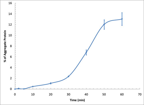
如图1所示,使用 PROTEOSTAT® 染料检测羊 lgG 在热诱导下的聚集情况,生成的曲线表明不同阶段 lgG 蛋白聚集的情况。

在刚开始阶段蛋白聚集情况较少,接着观察到生长阶段聚集体开始形成,最后生长率降低直至变成0,表明蛋白单体已几乎消失,差不多完全形成聚集体。


PROTEOSTAT® 蛋白聚集检测                              PROTEOSTAT® Protein aggregation assay

1:热诱导羊 IgG 蛋白聚集


监控机械诱导聚集


虽然普遍认为机械搅拌会加速蛋白聚集形成,但目前机械外力造成蛋白聚集形成的原理还不是十分明确。在药物研发中,可能受到抽真空或者过滤等产生的机械外力使蛋白药物形成聚集。

羊抗鼠 lgG(10 mg/mL)溶解在含有 10 mM 的磷酸钠,150 mM Nacl,0.05% 叠氮化钠,pH 7.2 的溶液中。机械搅拌的实验条件如下:室温、4 mL 的平底褐色玻璃瓶中加入 200 μL lgG 溶液,使用 Variomag® Poly electronic stirrer(2Mag-USA),以 990 rpm 速度进行搅拌。搅拌棒体积为1.0×0.4×0.2 cm3

在每个时间点,取10 μL lgG 用于检测。样品在 1×assay buffer 中稀释 10 倍后,得到的蛋白最终浓度为 1 mg/mL。在每个孔中加入 98 μL 的检测蛋白和 2 μL 的 PROTEOSTAT® Detection Reagent Loading Solution,每个时间点都做平行孔。

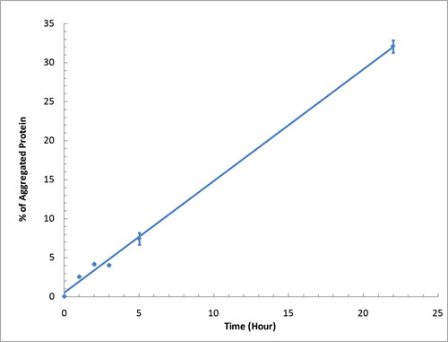
用 PROTEOSTAT® protein aggregation standards 作为标准曲线,定量检测机械搅拌诱导产生的蛋白聚集。在上述实验条件下,蛋白聚集与时间成正比例关系(图2)。


PROTEOSTAT® 蛋白聚集检测                              PROTEOSTAT® Protein aggregation assay

2:机械诱导样抗鼠 IgG 蛋白聚集


监控冷冻和反复冻融诱导聚集


冷冻对于蛋白稳定是至关重要的,因为在冷冻过程中会导致溶液的浓度发生变化。羊抗鼠 lgG(10 mg/mL)溶解在 10mM 的磷酸钠,150 mM Nacl,0.05% 叠氮化钠,pH 7.2 的溶液中。样品保存在 -20℃,反复冻融数次。

在每个时间点,取10 μL lgG 用于检测。样品在 1×assay buffer 中稀释 10 倍后,得到的蛋白最终浓度为 1 mg/mL。在每个孔中加入 98 μL 的检测蛋白和 2 μL 的 PROTEOSTAT® Detection Reagent Loading Solution,每个时间点做平行孔。

用 PROTEOSTAT® protein aggregation standards 作标准曲线,定量检测反复冻融诱导产生的蛋白聚集体。实验表明反复冻融也会诱导产生蛋白聚集。与热诱导引起蛋白聚集一样,会观察到一条S型曲线图。


PROTEOSTAT® 蛋白聚集检测                              PROTEOSTAT® Protein aggregation assay

产品编号 产品名称 产品规格 产品等级
ENZ-51023-KP050 PROTEOSTAT® Protein aggregation assay
 PROTEOSTAT ®  蛋白聚集检测
50 tests
ENZ-51023-KP002 PROTEOSTAT® Protein aggregation assay 2×96 tests

ProteoCarry <Protein Transfection Reagent> 无需预孵育!导入细胞质的蛋白转染试剂

  • 产品特性
  • 相关资料
  • Q&A
  • 参考文献

ProteoCarry <Protein Transfection Reagent>                              无需预孵育!导入细胞质的蛋白转染试剂

无需预孵育!导入细胞质的蛋白转染试剂                

ProteoCarry <Protein Transfection Reagent>

本产品是将蛋白以及葡聚糖等生物大分子导入细胞内,尤其是导入细胞质的试剂。传统的蛋白转染试剂存在蛋白滞留在内体或溶酶体中的问题,但是由于本产品可以高效地将蛋白运送至细胞质,可以广泛地适用于在评价导入蛋白的细胞内功能和抗体导入引起的功能抑制诱导等的实验。另外与传统产品不同的是,本品无需与蛋白进行预孵育,所以不会形成复合体,低限度地限制其对蛋白功能的影响。

※本产品仅供研究,研究以外请勿使用。


◆蛋白转染试剂及其问题


蛋白转染试剂(Proteofection试剂)是将重组蛋白和抗体直接导入细胞的工具。与将质粒导入以诱导表达目的蛋白的方法相比,可以更迅速地导入目的蛋白,因而受到了广泛地关注。

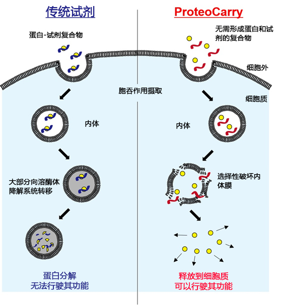
虽然已经开发了许多基于肽、阳离子的脂质或聚合物的蛋白转染试剂,但它们都是与蛋白形成复合物后,通过胞吞作用摄入细胞内,破坏内体膜将其运送至细胞质。然而,现有的大部分试剂内体膜的破坏活性低,并且从内体逃脱不足,导入的蛋白大部分都转移至溶酶体降解系统。本产品是一种新型肽蛋白转染试剂,克服了传统的蛋白转染试剂的缺点,通过强有力的内体膜选择性破坏活性,高效地向细胞质运送蛋白,使转导的蛋白在细胞内发挥活性。另外,与传统的蛋白转染试剂不同,本品无需与蛋白的复合物,不仅仅是蛋白,还可以向细胞质导入生物大分子(葡聚糖等)。

ProteoCarry <Protein Transfection Reagent>                              无需预孵育!导入细胞质的蛋白转染试剂

图1. 细胞导入原理的比较

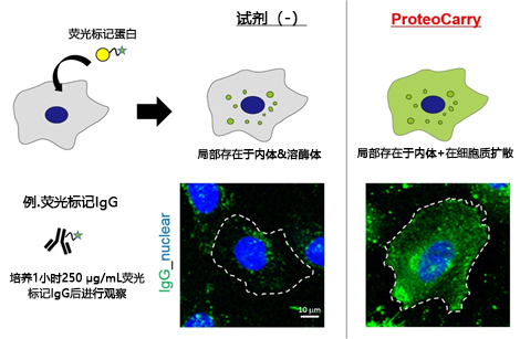
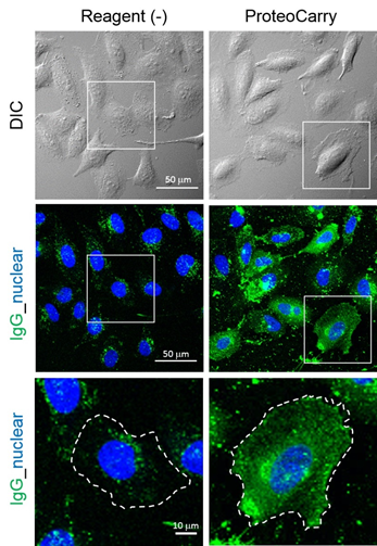
图 2 是导入荧光标记 IgG 的案例。不添加转染试剂时,以内体/溶酶体的染色为主,从细胞质中无法获得荧光信号,与之相对的,可以观察到在本产品案例中,内体、细胞质被大面积染色。(详情请参照应用实例)

ProteoCarry <Protein Transfection Reagent>                              无需预孵育!导入细胞质的蛋白转染试剂

图 2. 向 HeLa 细胞导入荧光标记 IgG 的比较


◆特点


● 基于肽特性的蛋白转染试剂,显示出高度水溶性。

● 优异的内体膜破坏活性,与传统方法相比显示出高细胞质传达性。

● 无需与导入物质进行预孵育,可以简便且迅速地转染。

● 由于不会与蛋白形成复合物,所以需要添加浓度较高的蛋白,但是可以低限度地抑制对蛋白功能的影响。

● 只需要1小时的处理就可以充分地转染至细胞质。

● 有无血清(<10% FBS)不会影响导入效率,可以根据想要使用的细胞设计实验。

● 已有向细胞质导入各种蛋白及生物大分子的应用实例。

● 例:IgG 抗体、功能性蛋白(Cre recombinase, Saponin)、多糖(葡聚糖)

● 推荐使用浓度几乎没有细胞毒性。


◆使用方法概述

ProteoCarry <Protein Transfection Reagent>                              无需预孵育!导入细胞质的蛋白转染试剂

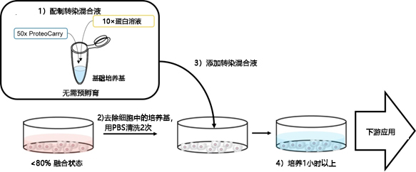
1. 向新鲜的培养基(无血清培养基,10% 以下含 FBS 培养基或 PBS)添加本产品以及想要导入的蛋白。(配制转染混合液)。

1. ※无需预孵育

2. 用 PBS 清洗 2 次细胞(80% 以下的融合状态)后,向细胞添加转染混合液。

3. 在转染混合液中培养细胞(~1 h)

4. 用 PBS 清洗 2 次细胞后,添加新鲜的培养基。

5. 实现各项应用。

◆试剂盒组成


● ProteoCarry(4 mg)

● FITC-dextran(2 mg,阳性对照用)

实验次数的预测


孔板的尺寸

ProteoCarry

FITC-dextran

6 well

14 assays

5 assays

12 well

28 assays

10 assays

24 well

56 assays

20 assays

48 well

140 assays

50 assays

96 well

280 assays

100 assays


◆有导入数据的细胞


HeLa, SW280, COS7, NIH3T3, HUVEC

参考导入量


导入物

推荐培养基的终浓度

抗体

50~250 μg/mL

蛋白

Saponin

1~10 μg/mL

Cre recombinase

10~100 μg/mL

FITC-dextran
    (阳性对照)

200 μg/mL


※导入效率根据细胞类型,细胞数量和蛋白类型各种因素改变。以上数据仅供参考,建议每次实验都要优化数据。

◆应用实例


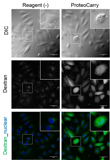
1. 向细胞内导入FITC-dextran


向 HeLa 细胞导入 FITC-Dextran(培养基的终浓度为 200 μg/mL),仅向细胞添加 FITC-Dextran 时,(Reagent(-))观察到点状的荧光信号,细胞质中几乎没有观察到信号;相反,使用本产品时扩散到了整个细胞质。


ProteoCarry <Protein Transfection Reagent>                              无需预孵育!导入细胞质的蛋白转染试剂


实验概要:


向 HeLa 细胞(80% confluent)添加 200 μg/mL FITC-dextran, 1×ProteoCarry in α-MEM,并培养 1 个小时。用 PBS 清洗细胞后,进行核染色(Hoechst)。


2. 向细胞内导入荧光标记 IgG


向 HeLa 细胞导入荧光标记 IgG 抗体(培养基终浓度为 250 μg/mL),仅有抗体添加至细胞时,可以观察到胞吞作用自发性摄入点状的荧光信号,表明抗体停留在内体和溶酶体中。另一方面,使用本产品时,不仅能观察到点状的内体结构,还可以观察到它主要在细胞质中广泛扩散。

※用荧光信号观察细胞扩散时,与内体相比,由于细胞质的体积较大,信号会被稀释。细胞质信号的检测需要添加高浓度的荧光标记蛋白,在本数据中添加了250 μg/mL的荧光标记 IgG。


ProteoCarry <Protein Transfection Reagent>                              无需预孵育!导入细胞质的蛋白转染试剂


实验概要:


向 HeLa 细胞(80% confluent)添加 250 μg/mL 荧光标记 IgG,1×ProteoCarry in α-MEM 并培养 1小时。用 PBS 清洗细胞后,进行核染色(Hoechst)。


3. 向细胞内导入 Cre recombinase


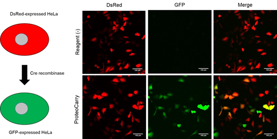
将设计为 Cre recombinase 不存在时表达 DsRed,只有在 Cre recombinase 存在时发生重组表达 GFP 的质粒向 HeLa 细胞导入基因。相对于导入第二天就表达 DsRed 的 HeLa 细胞,使用本产品导入 Cre recombinase(培养基终浓度为 10 μM),依赖导入细胞内的 Cre recombinase 表达 GFP,本产品在维持 Cre recombinase 的功能下将其导入了细胞。


ProteoCarry <Protein Transfection Reagent>                              无需预孵育!导入细胞质的蛋白转染试剂


实验概要:

向 HeLa 细胞转染质粒的第二天,向 HeLa 细胞添加 10 μM Cre recombinase,1×ProteoCarry in α-MEM,并培养 1 小时。用PBS清洗细胞后,用新的培养基再培养 24 小时。


◆保存


● 运输:室温

● 保存:-20℃


产品编号 产品名称 产品规格 产品等级
FDV-0015 ProteoCarry
 ProteoCarry <蛋白质转染试剂>
1 set

即用型彩色蛋白预染 Marker,稳定型 Protein MultiColor,Stable DynaMarker®

  • 产品特性
  • 相关资料
  • Q&A
  • 参考文献

即用型彩色蛋白预染 Marker,稳定型即用型彩色蛋白预染 Marker,稳定型                              Protein MultiColor,Stable DynaMarker®

Protein MultiColor,Stable DynaMarker®


在 4° C 下可以保存一年,即用型彩色蛋白预染 Marker,天然蛋白经预染标记。通过蛋白染色技术,实现高稳定性,可以保存于冰箱内。


即用型彩色蛋白预染 Marker,稳定型                              Protein MultiColor,Stable DynaMarker®


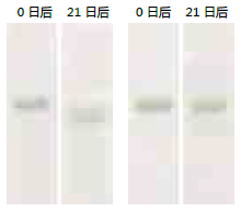
凝胶组成:5~20% 聚丙烯酰胺梯度凝胶,37°C孵育3个星期                


 染色 BSA


                                                             传统产品               本产品

即用型彩色蛋白预染 Marker,稳定型                              Protein MultiColor,Stable DynaMarker®

                                                          不同染色法下蛋白稳定性的比较


为了对比传统产品(DynaMarker® Protein MultiColorⅢ)以及使用本产品制备的染色蛋白(BSA)在 4° C 的稳定状态,先在 37° C 的严峻条件下孵育3个星期,然后再观察 SDS-PAGE 条带的状态。

本产品制备的染色 BSA,比传统产品更能保持条带的位置及形状,具有高稳定性。


产品编号 产品名称 产品规格 产品等级
DM660 Protein MultiColor, Stable, DynaMarker
即用型彩色蛋白预染Marker,稳定型
600 μL
DM660L Protein MultiColor, Stable, DynaMarker, Large
即用型彩色蛋白预染Marker,稳定型
5×600 μL

蛋白分子量标记

  • 产品特性
  • 相关资料
  • Q&A
  • 参考文献

蛋白分子量标记蛋白分子量标记


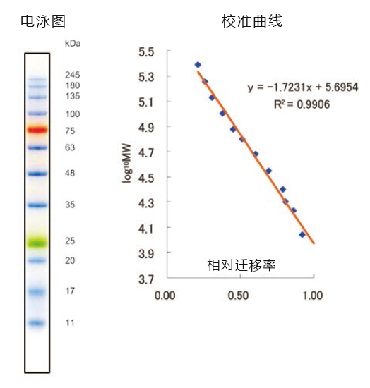
◆WIDE-VIEW™ 系列


该系列是预染蛋白SDS-PAGE用分子量标记。所有蛋白条带均为大肠杆菌表达的重组蛋白。所有的条带都与染料偶联,可用于在电泳中指示各个条带的位置。另外,本品还用作直接观察转膜效果。


■ 使用方法

将本产品恢复至室温并混匀,然后上样。


WIDE-VIEW™ Prestained Protein Size Marker Ⅲ

■ 产品规格及特点


   • 条带数:12

   • 分子量范围:11 – 245 kDa

   • 推荐使用量:5 µL/Lane

   • 无需稀释,煮沸

   • 带有用于电泳观察的颜色标记


蛋白分子量标记

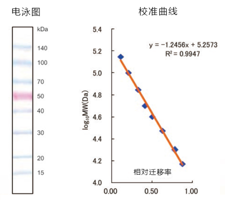
WIDE-VIEW™ Prestained Protein Size Marker

■ 产品规格及特点


   • 条带数:8

   • 分子量范围:15 – 140 kDa

   • 推荐使用量:5-10 µL/Lane

   • 无需稀释,煮沸


蛋白分子量标记

Western blotting 用标记

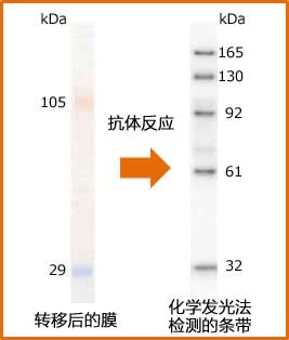
本产品是包含5种类型的抗体结合条带(32-165 kDa)和2种预染条带(蓝:29 kDa,橙:105 kDa)的 Western blotting 用蛋白分子量标记。种类型的抗体结合蛋白和后续所使用的抗体相结合,在检测过程中以条带显示,无需特殊处理或额外试剂。

所有蛋白条带均为大肠杆菌表达的重组蛋白。

■ 使用方法

将本产品恢复至室温并混匀,然后上样。

WESTERN-VIEW™ Western Protein Size Marker(32-165kDa)


■ 产品规格及特点


   • 条带数:8

   • 分子量范围:15 – 140 kDa

   • 推荐使用量:5-10 µL/Lane

   • 无需稀释,煮沸

   • 缓冲液成分:125 mmol/L Tris-HCl (pH6.8)、10 mmol/L DTT、5 mmol/L EDTA、17.4 w/v% glycerol、3 w/v% SDS、

         0.01 w/v% phenol red.

■ 注意


   • 请避免重复冻融。

   • 本产品的检测灵敏度与抗体和检测试剂密切相关。

   • 高灵敏度的检测系统中可能会出现额外的条带,请减少上样量。

   • 不适用于 Native-PAGE。


蛋白分子量标记


凝胶:SuperSep™Ace、10-20%,17 well

Running Buffer:SDS-PAGE缓冲液, pH 8.5

分子量标记:本产品(产品编号:236-02723) 5 μL/lane

膜:ClearTrans® SP的 PVDF 膜,疏水性,0.2 μm (产品编号:033-22453

抗体:抗山羊IgG,过氧化物酶标记

化学发光试剂:ImmunoStar® Zeta(产品编号:295-72404


◆产品列表


Prestained Marker

产品编号 产品名称 规格 包装
236-02463 WIDE-VIEW™ Prestained Protein Size Marker Ⅲ
    WIDE-VIEW™ 预染蛋白质标记Ⅲ
电泳用 25 μL
230-02461 500 μL
234-02464 500 μL×3
230-02221 WIDE-VIEW Prestained Protein Size Marker
    WIDE-VIEW 预染蛋白Marker
电泳用 500 μL

Western blotting 用标记

产品编号 产品名称 规格 包装
230-02721

WESTERN-VIEW Western Protein Size   Marker(32-165kDa)

WESTERN-VIEW™蛋白质标记(32-165kDa)

电泳用 25 μL
236-02723 250 μL

蛋白质大小标记物


产品编号

产品名称

规格

备注

230-02221

WIDE-VIEW™ Prestained

Protein Size MarkerⅢ

500 μL


已染色的蛋白质大小标记物。

各条带分子量:150, 100, 70,   50(pink), 40, 30, 20, 15(kDa)

294-63101

Molecular Weight Marker,   Low Range

1 mL

针对低分子的CBB染色用标记物。

各条带的分子量:42, 30, 20,   17, 6.5, 3.5(kDa)

131-14511

Molecular Weight Marker,   Middle Range

1 mL

针对中分子的CBB染色用标记物。

各条带的分子量:79, 42, 30,   20, 14(kDa)

296-63301

Molecular Weight Marker,   Wide Range

1 mL

CBB染色用的WideRange标记物。

各条带的分子量:180, 116,   97, 79, 42, 30, 20, 14, 6.5(kDa)

【参考链接】http://www.wako-chem.co.jp/siyaku/product/life/protein_marker/index.htm

产品编号 产品名称 产品规格 产品等级

WESTERN-VIEW™ Western蛋白marker WESTERN-VIEW™ Western Protein Size Marker(32-165kDa)

  • 产品特性
  • 相关资料
  • Q&A
  • 参考文献

WESTERN-VIEW™ Western 蛋白marker(32-165 kDa)WESTERN-VIEW™ Western蛋白marker                              WESTERN-VIEW™ Western Protein Size Marker(32-165kDa)


◆原理


      用于免疫印迹的蛋白marker,包括5种抗体结合的条带(32~165 kDa)和两种与色素体结合的条带(蓝色:29 kDa,橙色:105 kDa)。这 5种抗体结合的蛋白可以结合实验中使用的抗体,检测时会以条带形式出现,无需进行另外的实验步骤,也不需要另外的试剂。每种蛋白是E.coli表达的重组蛋白。可全程监控免疫印迹实验中蛋白的位置。


◆优点特色


  免疫印迹实验时可检测到

● 包括两种结合色素体的条带和5种抗体结合的条带

●  不需要煮沸或者稀释


◆案例应用


1、使用前将产品放置在室温

2、用枪头或则震荡仪充分混匀液体

3、每块胶 2~5 μL/道

4、转移到膜上后,在抗体反应时候,使用的抗体也会结合到marker上


WESTERN-VIEW™ Western蛋白marker                              WESTERN-VIEW™ Western Protein Size Marker(32-165kDa)

胶:SuperSep™ Ace, 10-20%, 17 wells

(产品编号:198-15041 

Running Buffer:SDS-PAGE Buffer, pH 8.5

(产品编号:192-16801


蛋白marker该产品(产品编号:236-02723 )5 μL/道


Clear Trans SP PVDF Membrane, Hydrophobic, 0.2 μm

(产品编号:033-22453 


抗体抗山羊IgG,过氧化氢标记

化学发光试剂 ImmunoStar Zeta(产品编号:295-72404


组成:

125 mmol/L Tris-HCl (pH 6.8), 10 mmol/L DTT, 5 mmol/L EDTA, 
17.4 w/v%
甘油, 3 w/v% SDS, 0.01 w/v% 酚红 

备注:

● 避免反复冻融

● 产品的检测灵敏度取决于抗体和使用的检测试剂

● 高灵敏度的检测系统会导致有额外的条带。请减少上样量。

● 与Native-PAGE实验不兼容

WESTERN-VIEW™ Western蛋白marker                              WESTERN-VIEW™ Western Protein Size Marker(32-165kDa)


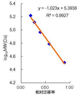
抗体结合条带(32~165 kDa)的标准曲线


产品列表

产品名称

规格

货号

级别

保存条件

WESTERN-VIEW™ Western Protein Size Marker(32 – 165 kDa)

25 μL(5~12次)

230-02721

电泳实验

-20℃

250 μL(50~120次)

236-02723


◆其他相关产品

产品名称

包装规格

货号

级别

保存条件

SuperSep™ Ace, 10-20%, 17 well

10块

198-15041

电泳

2~10℃

SDS-PAGE Buffer, pH 8.5

5 L

192-16801

25℃以下

ClearTrans SP PVDF Membrane, Hydrophobic

0.2 μM

033-22453

免疫印迹实验

室温

ImmunoStar Zeta

200 cm2

291-72401

-80℃

2000 cm2

295-72404

1000 cm2

297-72403


产品编号 产品名称 产品规格 产品等级
230-02721 WESTERN-VIEW™ Western Protein Size Marker(32-165kDa)
WESTERN-VIEW™ Western蛋白marker
25 μL
236-02723 WESTERN-VIEW™ Western Protein Size Marker(32-165kDa)
WESTERN-VIEW™ Western蛋白marker
25 μL

对照融合蛋白系列 重组蛋白纯化/检测用

  • 产品特性
  • 相关资料
  • Q&A
  • 参考文献

对照融合蛋白系列                              重组蛋白纯化/检测用

重组蛋白纯化/检测用

对照融合蛋白系列

对照融合蛋白系列                              重组蛋白纯化/检测用

对照融合蛋白系列                              重组蛋白纯化/检测用

◆Multitope蛋白


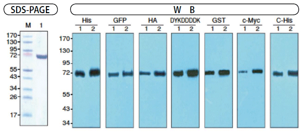
本品是由4种标签肽、3种标签蛋白连接成的重组蛋白。

因其为变性状态,可直接用于SDS-PAGE,在Western Blotting中可用作阳性对照。


对照融合蛋白系列                              重组蛋白纯化/检测用


通过各种一抗检测Multitope(Western Blotting检测法


对照融合蛋白系列                              重组蛋白纯化/检测用

Lane 1: MultiTope, 变性20ng
Lane 2: MultiTope, 变性50ng
 
<一抗>
His:6×His MoAb Albumine free, 5000倍稀释
GFP:Anti GFP(Wako, 012-22541), 1000倍稀释
HA:Anti HA(Wako, 014-21881), 500倍稀释
DYK:Anti DYKDDDDK(Wako, 018-22381), 

   5000倍稀释
GST:Anti GST(Wako, 013-21851), 500倍稀释
c-Myc:Anti c-Myc(Wako, 017-21871), 

    1000倍稀释
C端-His:Anti C-terminal 6×His

      (Wako, 010-21861), 400倍稀释


产品名称

备注

规格

产品编号

包装

Multitope蛋白

Multitope   (变性), 重组体, 溶液

Multitope, Denatured, recombinant, Solution

浓度:10 ng/μL

免疫化学用

139-16371

500 μL

133-16374

2.5 mL

  组成:50 mmol/L Tris-HCl, pH 6.8, 2 w/v% SDS, 10 v/v% glycerol, 0.1 w/v% bromophenol blue,

     1.5 w/v% DTT.
  使用方法:Load 5-10 μL of Multitope onto SDS-PAGE
  使用注意:本品保存温度为-20℃。
  保存过程中,缓冲液成分可能会析出,所以使用前请在室温条件下充分混合溶液直至析出成分完全溶解,

  方可使用。


◆DYKDDDDK-BAP, 重组体, 溶液


  本产品是可融合DYKDDDDK标签各个部位的Bacterial Alkaline Phosphatase(BAP)。


1.可用于识别抗DYKDDDDK标签抗体交叉性的

  对照蛋白。

2.可用于Western Blotting和免疫沉淀实验。

对照融合蛋白系列                              重组蛋白纯化/检测用


产品名称

备注

产品编号

包装

DYKDDDDK-BAP, 重组体, 溶液

N端 DYKDDDDK-BAP,重组体, 溶液

Amino-terminal DYKDDDDK-BAP, recombinant, Solution

基因研究用

016-24401

200 μg

N端 Met-DYKDDDDK-BAP, 重组体, 溶液

Amino-terminal Met-DYKDDDDK-BAP, recombinant, Solution

013-24411

200 μg

C端 DYKDDDDK-BAP, 重组体, 溶液

Carboxy-terminal DYKDDDDK-BAP, recombinant, Solution

036-22781

200 μg

 来源:大肠杆菌表达DYKDDDDK-BAP
 溶液组成: 10 mM Tris-HCl, pH8.0, 120 mM氯化钠, 0.05 mM氯化锌, 50 w/v %甘油
 蛋白浓度: 0.35 mg/mL
 含量: 90%以上(SDS-PAGE)
 分子量:

 约48.8 kDa (N端 DYKDDDDK-BAP)
 约49.0 kDa (N端 Met-DYKDDDDK-BAP)
 约48.8 kDa (C端 DYKDDDDK-BAP)


◆相关产品


内参照

下表为Western Blot实验中参照用单抗一览表。


产品名称

规格

产品编号

包装

抗β肌动蛋白单抗
  Anti β-Actin, Monoclonal Antibody

免疫化学用

013-24553

200 μg

011-24554

1 mg

017-24556

5 mg

抗β肌动蛋白单抗,过氧化物酶标记

Anti β-Actin, Monoclonal Antibody, Peroxidase Conjugated

免疫化学用

017-24573

200 μL

抗α-微管蛋白, 单抗

Anti α-Tubulin, Monoclonal Antibody

免疫化学用

011-25034

10 μg

017-25031

200 μg

013-25033

1 mg

  抗β-微管蛋白, 单抗

Anti β-Tubulin, Monoclonal Antibody

免疫化学用

018-25044

10 μg

014-25041

200 μg

010-25043

1 mg

  抗GAPDH, 单抗

Anti GAPDH, Monoclonal Antibody

免疫化学用

016-25523

200 μg

014-25524

1 mg

010-25526

5 mg

  抗GAPDH, 单抗, 过氧化酶结合

Anti GAPDH, Monoclonal Antibody, Peroxidase Conjugated

免疫化学用

015-25473

200 μL


蛋白酶抑制剂系列


产品名称

规格

产品编号

包装

蛋白酶抑制剂混合物试剂盒 I,无动物源成分(通用型)

Protease Inhibitor Cocktail Set I, Animal-derived-free (for general use) (×100), Lyophilized

通用型

165-26021

1 mL用

161-26023

1 mL用×5

蛋白酶抑制剂混合物试剂盒III,   DMSO溶液(不含EDTA)(×100)
  Protease Inhibitor Cocktail Set III, DMSO Solution   (EDTA free) (×100)

动物细胞

163-26061

1 mL

169-26063

1 mL×5

蛋白酶抑制剂混合物试剂盒V(无EDTA)(×100)
  Protease Inhibitor Cocktail Set V (EDTA free) (×100), Lyophilized

2D电泳

162-26031

1 mL

168-26033

1 mL×5

蛋白酶抑制剂混合物试剂盒 VI,DMSO溶液,用于植物(×100)
  Protease Inhibitor Cocktail Set VI, DMSO Solution (for Plant) (×100)

植物细胞

164-26091

1 mL

160-26093

1 mL×5

蛋白酶抑制剂混合物试剂盒VII,DMSO溶液,组氨酸标签蛋白(×100)
  Protease Inhibitor Cocktail Set VII, DMSO Solution (for Histidine-tagged Protein) (×100)

His Tag 纯化

167-26101

1 mL

163-26103

1 mL×5

※ 本页面产品仅供研究用,研究以外不可使用。

亲和标签比较表

亲和标签

TARGET

PA

6xHis

氨基酸序列

(YPGQ)5V

GVAMPGAEDDVV

HHHHHH

残基数

21

12

6

分子量 (kDa)

2.34

1.16

0.84

等电点(pI)

5.52

3.49

7.21

配体

TARGET tag

PA tag

Ni, Co, Zn, Cu

亲和珠回收

40% Propyleneglycol,
  1×TBS(pH7.5)

3M MgCl2, MES(pH6.0)

0.5M Imididazole(pH7.4)

单抗边界强度

(KD(M))

1.0×10⁻⁸

4.9×10⁻¹⁰

Ni-NTA:
  1.0×10⁻⁵~-6

单抗

Clone No.

P20.1(Mouse)

NZ-1(Rat)

Merck:HIS. H8(Mouse)

亲和力

++++

+++++

+++

提纯成本

+

+

+

多肽洗脱

+++

+++

Imidazole

背景

(培养上清)

+

+

+++++

背景

(细胞裂解物)

+

+

++++

亲和标签

DYKDDDDK

c-Myc

HA

GST

氨基酸序列

DYKDDDDK

EQKLISEEDL

YPYDVPDYA

残基数

8

10

9

218

分子量 (kDa)

1.01

1.20

1.10

26

等电点(pI)

3.97

4.00

3.56

配体

DYKDDDDK tag

c-Myc tag

HA tag

GST

亲和珠回收

Tris-HCl,
  Glycine Buffer(pH2~3.5)

Tris-HCl,
  Glycine Buffer(pH2~3.5)

Tris-HCl,
  Glycine Buffer(pH2~3.5)

Glutathione,
  Tris-HCl,
  Glycine Buffer(pH2~3.5)

单抗边界强度

(KD(M))

2.8×10⁻⁸

2.2×10⁻⁹

2.8×10⁻⁹

Glutathione:
  1.0×10⁻⁵~-6

单抗

Clone No.

M2(Mouse)

9E10(Mouse)

3F10(Mouse)
  HA-7(Rat)

Wako:5A7(Mouse)

亲和力

++++

++++

++++

++++

提纯成本

++

++

++

+

多肽洗脱

+++

++

++

Glutathione

背景

(培养上清)

++++

+++

+++

++++

背景

(细胞裂解物)

++++

++++

+++

++++

  本比较表为Wako独家调查的数据。样品、检测方法和手法的不同,特异性也会有所变化,因此无法保证各个亲和标签的功能。


产品编号 产品名称 产品规格 产品等级

亲和标签抗体结合磁珠 重组蛋白纯化用

  • 产品特性
  • 相关资料
  • Q&A
  • 参考文献

亲和标签抗体结合磁珠                              重组蛋白纯化用

重组蛋白纯化用

亲和标签抗体结合磁珠

亲和标签抗体结合磁珠                              重组蛋白纯化用

亲和标签抗体结合磁珠                              重组蛋白纯化用

◆抗DYKDDDDK tag抗体磁珠


  已固定识别DYKDDDDK肽单抗的磁珠悬浮液。可用于检测大肠杆菌、酵母、哺乳动物表达的DYKDDDDK标签融合蛋白。


特点


● 非特异性结合少

● 可回收N端、C端、Internal全部蛋白

● 可用DYKDDDDK肽、SDS溶液洗脱

● 无需离心处理,可减少样品损耗,简单替换溶液


1. 抗DYKDDDDK标签抗体磁珠的性能

亲和标签抗体结合磁珠                              重组蛋白纯化用
可在不依赖标签位置的情况下,回收重组蛋白。

把各DYKDDDDK tag融合蛋白分别加入细胞溶解液中进行免疫沉淀后,通过Western blotting比较抗原回收量。

经4%SDS样品缓冲液洗脱和SDS-PAGE后,用Western blotting法,加过氧化酶标签抗体(产品编号:015-22391)进行发光检测。
 
M : 标签蛋白
Met : N-terminal Met-DYKDDDDK-BAP
N : N-terminal DYKDDDDK-BAP
C : C-terminal DYKDDDDK-BAP

2. 洗脱法比较抗原回收性能

亲和标签抗体结合磁珠                              重组蛋白纯化用
 SDS和DYKDDDDK肽均能洗脱抗原

将DYKDDDDK标签融合蛋白加入细胞溶解液中,分别加入磁珠吸附后,利用各种洗脱法回收抗原量并用Western Blotting法进行比较。

用过氧化酶标签抗体(产品编号:015-22391)进行发光检测。
 
M:Marker蛋白
Lane1添加2×SDS sample buffer后,煮沸
Lane2DYKDDDDK peptide(200 μg/mL)
Lane34% SDS solution

3. 非特异性吸附蛋白的比较


亲和标签抗体结合磁珠                              重组蛋白纯化用

在不含DYKDDDDK tag的细胞溶解液中分别添加磁珠并进行免疫沉淀后,比较非特异性吸附蛋白。

用4%SDS样品缓冲液洗脱、电泳后,用银染色、抗小鼠IgG、过氧化酶标签抗体进行Western Blotting比较。

无法检测出非特异性吸附质。


产品名称

子类
  克隆号

规格

产品编号

包装

DYK标签:DYKDDDK

抗DYKDDDDKtag抗体磁珠

Anti DYKDDDDK tag Magnetic Beads

小鼠 IgG2b

1E6

免疫化学用

011-25154

200 μL

017-25151

2.5 mL

013-25153

2.5 mL×5

 溶液组成:10 mg/mL Anti-DYKDDDDK Magnetic Beads, 1×TBS(pH 7.4), 50 w/v % glycerol, 0.05 w/v %       sodium azide


◆相关产品


内参照

下表为Western Blot实验中参照用单抗一览表。


产品名称

规格

产品编号

包装

抗β肌动蛋白单抗
  Anti β-Actin, Monoclonal Antibody

免疫化学用

013-24553

200 μg

011-24554

1 mg

017-24556

5 mg

抗β肌动蛋白单抗,过氧化物酶标记

Anti β-Actin, Monoclonal Antibody,   Peroxidase Conjugated

免疫化学用

017-24573

200 μL

抗α-微管蛋白, 单抗

Anti α-Tubulin, Monoclonal Antibody

免疫化学用

011-25034

10 μg

017-25031

200 μg

013-25033

1 mg

Anti β-Tubulin, Monoclonal Antibody
  抗β-微管蛋白, 单抗

免疫化学用

018-25044

10 μg

014-25041

200 μg

010-25043

1 mg

Anti GAPDH, Monoclonal Antibody
  抗GAPDH, 单抗

免疫化学用

016-25523

200 μg

014-25524

1 mg

010-25526

5 mg

Anti GAPDH, Monoclonal Antibody, Peroxidase   Conjugated
  抗GAPDH, 单抗, 过氧化酶结合

免疫化学用

015-25473

200 μL


蛋白酶抑制剂系列


产品名称

规格

产品编号

包装

蛋白酶抑制剂混合物试剂盒 I,无动物源成分(通用型)

Protease Inhibitor Cocktail Set I, Animal-derived-free (for general use) (×100), Lyophilized

通用型

165-26021

1 mL用

161-26023

1 mL用×5

蛋白酶抑制剂混合物试剂盒III,   DMSO溶液(不含EDTA)(×100)
  Protease Inhibitor Cocktail Set III, DMSO Solution   (EDTA free) (×100)

动物细胞

163-26061

1 mL

169-26063

1 mL×5

蛋白酶抑制剂混合物试剂盒V (无EDTA)(×100)
  Protease Inhibitor Cocktail Set V (EDTA free) (×100), Lyophilized

2D电泳

162-26031

1 mL

168-26033

1 mL×5

蛋白酶抑制剂混合物试剂盒 VI,DMSO溶液,用于植物(×100)
  Protease Inhibitor Cocktail Set VI, DMSO Solution (for Plant) (×100)

植物细胞

164-26091

1 mL

160-26093

1 mL×5

蛋白酶抑制剂混合物试剂盒VII,DMSO溶液,组氨酸标签蛋白(×100)
  Protease Inhibitor Cocktail Set VII, DMSO Solution   (for Histidine-tagged Protein) (×100)

His Tag 纯化

167-26101

1 mL

163-26103

1 mL×5

※ 本页面产品仅供研究用,研究以外不可使用。


亲和标签比较表

亲和标签

TARGET

PA

6xHis

氨基酸序列

(YPGQ)5V

GVAMPGAEDDVV

HHHHHH

残基数

21

12

6

分子量 (kDa)

2.34

1.16

0.84

等电点(pI)

5.52

3.49

7.21

配体

TARGET tag

PA tag

Ni, Co, Zn, Cu

亲和珠回收

40% Propyleneglycol,
  1×TBS(pH7.5)

3M MgCl2, MES(pH6.0)

0.5M Imididazole(pH7.4)

单抗边界强度

(KD(M))

1.0×10⁻⁸

4.9×10⁻¹⁰

Ni-NTA:
  1.0×10⁻⁵~-6

单抗

Clone No.

P20.1(Mouse)

NZ-1(Rat)

Merck:HIS. H8(Mouse)

亲和力

++++

+++++

+++

提纯成本

+

+

+

多肽洗脱

+++

+++

Imidazole

背景

(培养上清)

+

+

+++++

背景

(细胞裂解物)

+

+

++++

亲和标签

DYKDDDDK

c-Myc

HA

GST

氨基酸序列

DYKDDDDK

EQKLISEEDL

YPYDVPDYA

残基数

8

10

9

218

分子量 (kDa)

1.01

1.20

1.10

26

等电点(pI)

3.97

4.00

3.56

配体

DYKDDDDK tag

c-Myc tag

HA tag

GST

亲和珠回收

Tris-HCl,
  Glycine Buffer(pH2~3.5)

Tris-HCl,
  Glycine Buffer(pH2~3.5)

Tris-HCl,
  Glycine Buffer(pH2~3.5)

Glutathione,
  Tris-HCl,
  Glycine Buffer(pH2~3.5)

单抗边界强度

(KD(M))

2.8×10⁻⁸

2.2×10⁻⁹

2.8×10⁻⁹

Glutathione:
  1.0×10⁻⁵~-6

单抗

Clone No.

M2(Mouse)

9E10(Mouse)

3F10(Mouse)
  HA-7(Rat)

Wako:5A7(Mouse)

亲和力

++++

++++

++++

++++

提纯成本

++

++

++

+

多肽洗脱

+++

++

++

Glutathione

背景

(培养上清)

++++

+++

+++

++++

背景

(细胞裂解物)

++++

++++

+++

++++

  本比较表为Wako独家调查的数据。样品、检测方法和手法的不同,特异性也会有所变化,因此无法保证各个亲和标签的功能。


产品编号 产品名称 产品规格 产品等级

亲和标签抗体系列 重组蛋白纯化检测用

  • 产品特性
  • 相关资料
  • Q&A
  • 参考文献

亲和标签抗体系列                              重组蛋白纯化检测用

重组蛋白纯化检测用

亲和标签抗体系列

亲和标签抗体系列                              重组蛋白纯化检测用

亲和标签抗体系列                              重组蛋白纯化检测用

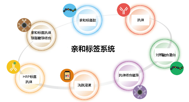
  和光为您准备了DYKDDDDK, 6×His, GFP, HA, MBP, c-Myc, GST,V5融合蛋白提纯、检测用抗体及珠。
  另外,和光还准备了上述各样品的试用装,您可向当地代理商索取。


◆DYK标签:DYKDDDDK


  DYK标签是识别DYKDDDDK肽的单抗。可用于检测大肠杆菌、酵母、哺乳动物中的DYKDDDDK标签融合蛋白。


亲和标签抗体系列                              重组蛋白纯化检测用


数据提供: 
鹿儿岛大学研究生院医疗牙科综合研究科 附属顽固病毒病理控制研究中心 分子病毒感染研究领域 草野秀一老师

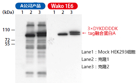
将表达3×DYKDDDDK标签融合蛋白A的质粒转染至HEK293细胞株中,经过24小时培养后,随机选取其中的克隆细胞。将选取的克隆细胞用RIPA缓冲液进行溶解,对所得的细胞溶解液用Western Blot法检测,可检测出3×FLAG标签融合蛋白。

分别用Wako(克隆号 1E6)(产品编号:018-22381)抗体和A公司抗体进行Western Blot检测。

与A公司抗体相比,Wako(克隆号 1E6)抗体识别DYKDDDDK的特异性更高。


A公司产品和Wako产品在Western Blot检测中的灵敏度比较


亲和标签抗体系列                              重组蛋白纯化检测用

Lane 1: 293T细胞溶解液
Lane 2: 瞬时表达蛋白A-DYKDDDDK的293T细胞溶解液
Lane 3: 瞬时表达蛋白B-DYKDDDDK的293T細胞溶解液
封闭:含5%脱脂奶的TBS-T、4℃,一晚

抗体反应:2小时、室温
发光剂:ImmunoStar® LD
检测:LAS-3000、曝光时间15分

Wako 1E6-HRP比A公司产品有更高的灵敏度!!


产品名称

子类克隆号

适用实验

规格

产品编号

包装

单抗

抗DYKDDDDK标签,单抗

Anti DYKDDDDK tag, Monoclonal Antibody

小鼠IgG2b   1E6

WB,IP,FCM,ChIP

免疫化学用

018-22381

200 μg

014-22383

1 mg

012-22384

5 mg

抗DYKDDDDK标签,单抗

Anti DYKDDDDK tag,Monoclonal Antibody,Rat

大鼠IgG1   6F7

WB,IP,ELISA,IHC,FCM

免疫化学用

018-23621

200 μg

012-23624

1 mg

HRP标签单抗

抗DYKDDDDK标签,单抗,过氧化酶结合

Anti DYKDDDDK tag, Monoclonal Antibody, Peroxidase Conjugated

小鼠IgG2b   1E6

WB

免疫化学用

015-22391

200 μL

019-22394

1 mL

◆6×His标签:HHHHHH (6×组氨酸)


  可用于检测His(6×组氨酸)标签融合蛋白。克隆不同,识别端的特异性、应用也有所不同。


亲和标签抗体系列                              重组蛋白纯化检测用

克隆号

识别端

Western blot

Immunoprecipitation

ELISA

产品编号

N端

C端

N端

C端

9F2

C端

ND

+++++

ND

++

010-21861

9C11

N・C端

+++++

++++

+++++

++

011-23091

21-48

N・C端

+++

++++

+++

+++

017-23211

28-75

N・C端

++

++

+++++

+++++

014-23221

9F2

C端

ND

+++++

013-23171

9C11

N・C端

+++++

++++

010-23181

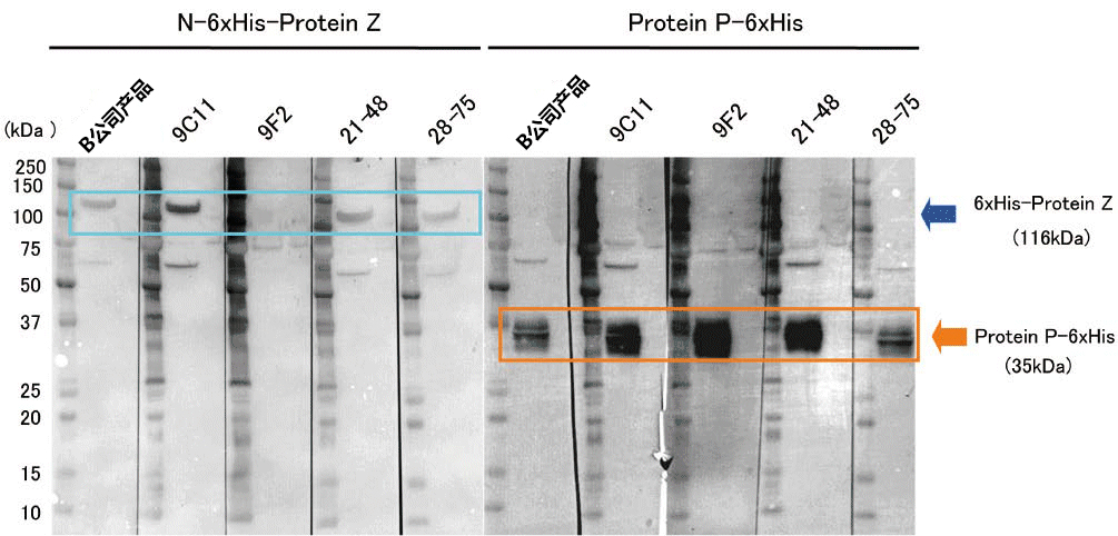
B公司产品和Wako产品各克隆号的Western blot检测功能比较


亲和标签抗体系列                              重组蛋白纯化检测用

N端His:HEK293T表达的6×His-

ProteinZ, 1×10⁵ cells
C
端His:Protein P-6×His+CHO细胞溶解液, 1×10⁵ cells

一抗:1 μg/mL
二抗:抗小鼠IgG抗体, 5000倍稀释液
可确认其具有比B公司产品同等甚至更高的
灵敏度。


6×His tag抗体 选购指南


  • N端的检测中,最强的克隆号是什么?
    WB:9C11 IP: 9C11, 28-75

  • C端的检测中,最强的克隆号是什么
    WB:9F2 IP:28-75

  • 适用于WB检测中,识别N/C端的克隆号是什么?
    9C11

  • 适用于IP检测中,识别N/C端的克隆号是什么?
    28-75

  • 可灵活应用于WB/IP的万能型克隆号是哪个?
    21-48

  • 适用于WB检测的克隆号是哪个?
    9C11

  • 适用于IP检测的克隆号是哪个?
    28-75

  • 只识别C端的性价比较高的克隆号是哪个?
    9F2


产品名称

子类

克隆号

适用实验

规格

产品编号

包装

单抗

抗6×组氨酸,单抗

Anti 6×His,Monoclonal Antibody(9F2)

小鼠IgG1
  9F2

WB,IP

免疫化学用

010-21861

200 μg

014-21864

1 mg

010-21866

5 mg

抗6×组氨酸,单抗

Anti 6×His,Monoclonal Antibody(9C11)

小鼠IgG1   9C11

WB,IP,ELISA

免疫化学用

011-23091

200 μg

015-23094

1 mg

011-23096

5 mg

抗6×组氨酸,单抗

Anti 6×His,Monoclonal Antibody(21-28)

小鼠IgG1・κ
  21-48

WB,IP,ELISA

免疫化学用

017-23211

200 μg

011-23214

1 mg

抗6×组氨酸,单抗

Anti 6×His,Monoclonal Antibody(28-75)

小鼠IgG3・κ
  28-75

WB,IP,ELISA

免疫化学用

014-23221

200 μg

018-23224

1 mg

HRP标签单抗

抗6×组氨酸,单抗,过氧化酶结合

Anti 6×Histidine, Monoclonal Antibody(9F2), Peroxidase Conjugated

小鼠IgG1   9F2

WB

免疫化学用

013-23171

100 μL

017-23174

500 μL

抗6×组氨酸,单抗,过氧化酶结合

Anti 6×Histidine, Monoclonal Antibody(9C11), Peroxidase   Conjugated

小鼠IgG1   9C11

WB

免疫化学用

010-23181

100 μL

014-23184

500 μL

◆GFP tag: Green Fluorescent Protein (GFP)


  可识别Green Fluorescent Protein (GFP)抗体。GFP是可实时检测目标蛋白局部或细胞、细胞器中的标签蛋白,可应用于各个领域。


产品名称

子类
  克隆号

适用实验

规格

产品编号

包装

单抗及多抗

抗绿色荧光蛋白,单抗

Anti Green Fluorescent Protein, Monoclonal Antibody (Anti GFP, MoAb)

小鼠IgG2a・κ
  mFX73

IP, IHC

免疫化学用

012-20461

100 μL

018-20463

100 μL×5

抗绿色荧光蛋白,单抗

Anti GFP,MoAb(mF×75)

小鼠IgG2a・κ
  mFX75

WB

免疫化学用

012-22541

100 μL

抗绿色荧光蛋白,多抗 IS,兔

Anti Green Fluorescent Protein,Rabbit Polyclonal IS

WB, IHC

免疫化学用

013-23811

20 μg

◆HA tag:YPYDVPDYA


  血凝素(HA)来源(98-106 aa)的多肽:抗YPYDVPDYA单抗。


产品名称

子类
  克隆号

适用

实验

规格

产品编号

容量

单抗

抗血凝素, 单抗

Anti HA,MoAb

小鼠IgG2b   4B2

WB, IP

免疫化学用

014-21881

200 μg

018-21884

1 mg

014-21886

5 mg

HRP标签单抗

抗血凝素, 单抗,过氧化酶结合

Anti Hemagglutinin,Monoclonal Antibody, Peroxidase   Conjugated

小鼠IgG2b   4B2

WB

免疫化学用

011-21911

100 μL

015-21914

500 μL


参考数据


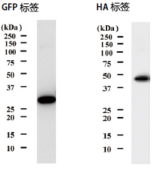
 抗绿色荧光蛋白, 单抗(012-22541
 抗体浓度:1 μg/mL
 抗原量:将100 μL表达GFP重组蛋白的大肠杆菌溶解液进行10倍稀释,并取

    10 μL进行电泳操作
 用10000倍稀释的抗小鼠IgG-HRP抗体进行检测。
 
 抗血凝素,单抗(
014-21881

 抗体浓度:0.5 μg/mL
 抗原量:对5 ng HA标签重组蛋白进行电泳操作
 用10000倍稀释的抗小鼠IgG-HRP抗体进行检测。  

亲和标签抗体系列                              重组蛋白纯化检测用

◆MBP tag:Maltose Binding Protein


  可识别Maltose-binding Protein (MBP)单抗。MBP是对麦芽糖、麦芽糊精具有结合性的蛋白,具有通过与目的蛋白融合,提高表达蛋白可溶性的特点,可简便纯化、检测重组蛋白。


产品名称

子类
  克隆号

适用实验

规格

产品编号

包装

单抗

抗麦芽糖结合蛋白,单抗

Anti MBP, Monoclonal Antibody,   Rat

大鼠IgG2b

TMab-2

ELISA, WB

免疫化学用

016-24141

200 μg

◆c-Myc标签:EQKLISEEDL


  可识别人c-Myc 来源(410-419aa)多肽EQKLISEEDL单抗。


产品名称

子类
  克隆号

适用实验

规格

产品编号

包装

单抗

抗c-Myc,单抗

ANTI c-Myc, Monoclonal Antibody

小鼠IgG1

9E10

WB, IP

免疫化学用

017-21871

200 μg

011-21874

1 mg

017-21876

5 mg

抗c-Myc,单抗

Anti c-Myc, Monoclonal Antibody (10D11)

小鼠IgG1

10D11

WB, IP

免疫化学用

017-26511

20 μg

013-26513

200 μg

011-26514

1 mg

HRP标签单抗

抗c-Myc, 单抗,过氧化酶结合

Anti c-Myc,MoAb, Peroxidase Conjugated

小鼠IgG1

9E10

WB

免疫化学用

014-21901

100 μL

018-21904

500 μL

抗c-Myc, 单抗,过氧化酶结合

Anti c-Myc, Monoclonal Antibody, Peroxidase Conjugated (10D11)

小鼠IgG1

10D11

WB

免疫化学用

014-26521

10 μL

010-26523

100μL

018-26524

500 μL


◆GST标签:Glutathione-S-transferase


  可识别Glutathione S-transferase(GST)单抗。


产品名称

子类
  克隆号

适用实验

规格

产品编号

包装

单抗

抗谷胱甘肽 S-转移酶,单抗

Anti Glutathione S-transferase, Monoclonal Antibody

小鼠IgG2b
  5A7

WB, IP

免疫化学用

013-21851

200 μg

017-21854

1 mg

013-21856

5 mg

HRP标签单抗

抗谷胱甘肽 S-转移酶,单抗,过氧化酶结合

Anti Glutathione S-transferse,Monoclonal Antibody, HRP Conjugated

小鼠IgG2b   5A7

WB

免疫化学用

011-21891

100 μL

015-21894

500 μL


参考数据


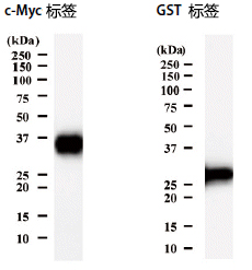
抗c-Myc, 单抗(017-21871
抗体浓度:0.25 μg/mL
抗原量:对10 ng c-Myc标签重组蛋白进行电泳操作。
用10000倍稀释的抗小鼠IgG-HRP抗体进行检测。
 
 抗谷胱甘肽 S-转移酶, 单抗(
013-21851
 抗体浓度:1.0 μg/mL
 抗原量:将100 μL表达GTS标签重组蛋白的大肠杆菌溶解液进行10倍稀释,

     并取10 μL进行电泳操作。
  用10000倍稀释的抗小鼠IgG-HRP抗体进行检测。

亲和标签抗体系列                              重组蛋白纯化检测用

◆V5 tag:GKPIPNPLLGLDST


  可识别多肽GKPIPNPLLGLDST单抗。


产品名称

子类
  克隆号

适用实验

规格

产品编号

包装

单抗

抗V5 tag, 单抗

Anti V5 tag, Monoclonal Antibody(6F5)

小鼠IgG2a
  6F5

WB, IP

免疫化学用

011-23591

200 μg

015-23594

1 mg

011-23596

5 mg

HRP标签单抗

抗V5tag,单抗,过氧化酶结合

Anti V5 tag, Monoclonal Antibody(6F5), Peroxidase Conjugated

小鼠IgG2a

6F5

WB

免疫化学用

019-24371

100 μL


◆相关产品


内参照

下表为Western Blot实验中参照用单抗一览表。


产品名称

规格

产品编号

包装

抗β肌动蛋白单抗
  Anti β-Actin, Monoclonal Antibody

免疫化学用

013-24553

200 μg

011-24554

1 mg

017-24556

5 mg

抗β肌动蛋白单抗,过氧化物酶标记

Anti β-Actin, Monoclonal Antibody, Peroxidase Conjugated

免疫化学用

017-24573

200 μL

抗α-微管蛋白,单抗

Anti α-Tubulin, Monoclonal Antibody

免疫化学用

011-25034

10 μg

017-25031

200 μg

013-25033

1 mg

抗β-微管蛋白,单抗

Anti β-Tubulin, Monoclonal Antibody

免疫化学用

018-25044

10 μg

014-25041

200 μg

010-25043

1 mg

抗GAPDH,单抗

Anti GAPDH, Monoclonal Antibody

免疫化学用

016-25523

200 μg

014-25524

1 mg

010-25526

5 mg

  抗GAPDH,单抗,过氧化酶结合

Anti GAPDH, Monoclonal Antibody, Peroxidase Conjugated

免疫化学用

015-25473

200 μL


蛋白酶抑制剂系列


产品名称

规格

产品编号

包装

蛋白酶抑制剂混合物试剂盒 I,无动物源成分(通用型)

Protease Inhibitor Cocktail Set I, Animal-derived-free (for general use) (×100), Lyophilized

通用型

165-26021

1 mL用

161-26023

1 mL用×5

蛋白酶抑制剂混合物试剂盒III,DMSO溶液(不含EDTA)(×100)
  Protease Inhibitor Cocktail Set III, DMSO Solution (EDTA free) (×100)

动物细胞

163-26061

1 mL

169-26063

1 mL×5

蛋白酶抑制剂混合物试剂盒V(无EDTA)(×100)
  Protease Inhibitor Cocktail Set V (EDTA free) (×100), Lyophilized

2D电泳

162-26031

1 mL

168-26033

1 mL×5

蛋白酶抑制剂混合物试剂盒 VI,DMSO溶液,用于植物(×100)
  Protease Inhibitor Cocktail Set VI, DMSO Solution (for Plant) (×100)

植物细胞

164-26091

1 mL

160-26093

1 mL×5

蛋白酶抑制剂混合物试剂盒VII,DMSO溶液,组氨酸标签蛋白(×100)
  Protease Inhibitor Cocktail Set VII, DMSO Solution (for Histidine-tagged Protein) (×100)

His Tag 纯化

167-26101

1 mL

163-26103

1 mL×5

※ 本页面产品仅供研究用,研究以外不可使用。


亲和标签比较表

亲和标签

TARGET

PA

6xHis

氨基酸序列

(YPGQ)5V

GVAMPGAEDDVV

HHHHHH

残基数

21

12

6

分子量 (kDa)

2.34

1.16

0.84

等电点(pI)

5.52

3.49

7.21

配体

TARGET tag

PA tag

Ni, Co, Zn, Cu

亲和珠回收

40% Propyleneglycol,
  1×TBS(pH7.5)

3M MgCl2, MES(pH6.0)

0.5M Imididazole(pH7.4)

单抗边界强度

(KD(M))

1.0×10⁻⁸

4.9×10⁻¹⁰

Ni-NTA:
  1.0×10⁻⁵~-6

单抗

Clone No.

P20.1(Mouse)

NZ-1(Rat)

Merck:HIS. H8(Mouse)

亲和力

++++

+++++

+++

提纯成本

+

+

+

多肽洗脱

+++

+++

Imidazole

背景

(培养上清)

+

+

+++++

背景

(细胞裂解物)

+

+

++++

亲和标签

DYKDDDDK

c-Myc

HA

GST

氨基酸序列

DYKDDDDK

EQKLISEEDL

YPYDVPDYA

残基数

8

10

9

218

分子量 (kDa)

1.01

1.20

1.10

26

等电点(pI)

3.97

4.00

3.56

配体

DYKDDDDK tag

c-Myc tag

HA tag

GST

亲和珠回收

Tris-HCl,
  Glycine Buffer(pH2~3.5)

Tris-HCl,
  Glycine Buffer(pH2~3.5)

Tris-HCl,
  Glycine Buffer(pH2~3.5)

Glutathione,
  Tris-HCl,
  Glycine Buffer(pH2~3.5)

单抗边界强度

(KD(M))

2.8×10⁻⁸

2.2×10⁻⁹

2.8×10⁻⁹

Glutathione:
  1.0×10⁻⁵~-6

单抗

Clone No.

M2(Mouse)

9E10(Mouse)

3F10(Mouse)
  HA-7(Rat)

Wako:5A7(Mouse)

亲和力

++++

++++

++++

++++

提纯成本

++

++

++

+

多肽洗脱

+++

++

++

Glutathione

背景

(培养上清)

++++

+++

+++

++++

背景

(细胞裂解物)

++++

++++

+++

++++

  本比较表为Wako独家调查的数据。样品、检测方法和手法的不同,特异性也会有所变化,因此无法保证各个亲和标签的功能。


产品编号 产品名称 产品规格 产品等级

Ni-NTA琼脂糖 用于蛋白提取

  • 产品特性
  • 相关资料
  • Q&A
  • 参考文献

用于蛋白提取Ni-NTA琼脂糖                              用于蛋白提取

Ni-NTA琼脂糖


  本产品可用于亲和提取在N末端或C末端融合了带有六个组氨酸残基的6×His标签的重组蛋白。它用于提取多种蛋白质,如可溶性蛋白质,不溶性蛋白质,膜蛋白质等等。溶出时无需使用多肽,以低成本和简单的操作即可提取目的蛋白。


◆特点


● 具有高性价比

● 可再生利用


◆产品规格

 凝胶基质

 6% 交联琼脂糖

 配体

 次氮基三乙酸(NTA)

 Beads size

 50-150 μm

 蛋白结合容量

 >50 mg/mL 凝胶

*蛋白结合容量会因目的蛋白不同而有所差异。


数据


Ni-NTA琼脂糖                              用于蛋白提取


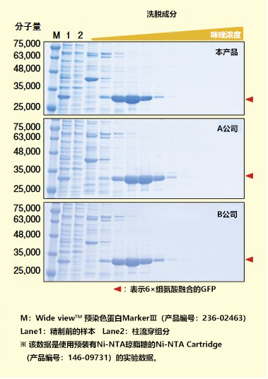
可以与其他公司的产品回收等量的蛋白质。


<实验条件>

① 通过超声破碎法从大肠杆菌中回收6×组氨酸融合GFP,作为精制前样本。

② 将Ni–NTA柱连接到液相色谱系统。

③ 用150 mL Running Buffer*1平衡色谱柱。

④ 上样精制前样本。

⑤ 用100 mL Running Buffer清洗。

⑥ 用150 mL Elution Buffer*2洗脱。

⑦ 用100 mL Running Buffer清洗。

⑧ 进行SDS-PAGE、CBB染色。


*1 Running Buffer:20 mmol/L Sodium Phosphate(pH 7.4),

          500 mmol/L Sodium Chloride, 

          10 mmol/L Imidazole

*2 Elution Buffer:20 mmol/L Sodium Phosphate(pH 7.4),

           500 mmol/L Sodium Chloride,

                                10~500 mmol/L Imidazole


具体实验条件请根据精制的目的蛋白进行检讨。


◆产品列表

产品编号

产品名称

规格

包装

147-09761

Ni-NTA Agarose
Ni-NTA琼脂糖

基因研究用

2 mL(Net 1 mL)

143-09763

10 mL(Net 5 mL)

141-09764

100 mL(Net 50 mL)


相关产品


预装型


Ni-NTA琼脂糖                              用于蛋白提取


本产品是预装柱产品。可直接用于液相色谱系统。

产品编号

产品名称

规格

包装

146-09731

Ni-NTA Cartridge
Ni-NTA预装柱

基因研究用

1EA(5 mL)

142-09733

1EA(5 mL)×5


高性能型


具有高蛋白结合能力的高性能型Ni-NTA琼脂糖。

产品编号

产品名称

规格

包装

149-09684

Ni-NTA Agarose HP
Ni-NTA琼脂糖 高性能型

基因研究用

2 mL(Net 1 mL)

145-09681

10 mL(Net 5 mL)

141-09683

100 mL(Net 50 mL)

※ 本页面产品仅供研究用,研究以外不可使用。

产品编号 产品名称 产品规格 产品等级

PA Tag 新型标签系统 PA Tag

  • 产品特性
  • 相关资料
  • Q&A
  • 参考文献

PA tagPA Tag 新型标签系统                              PA Tag

亲和纯化标签蛋白!

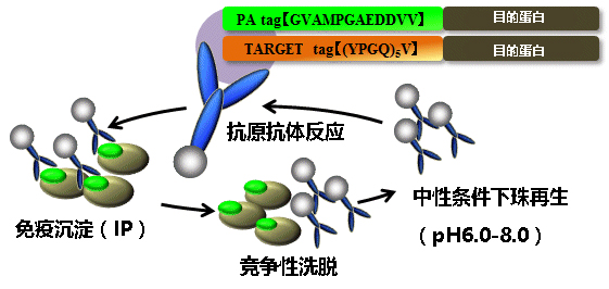
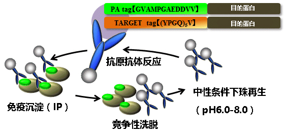
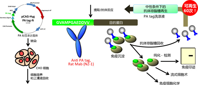
  PA tag 系统是利用高亲和性&高特异性单抗(NE-1)识别肽标签(GVAMPGAEDDVV)的重组蛋白新型检测系统。PA tag 系统中的抗体琼脂糖可通过免疫沉淀法纯化 PA tag 融合蛋白,并可在洗涤后获得再生。中性 pH 条件下 PA tag 肽简单方便地竞争性洗脱 PA tag 融合蛋白。

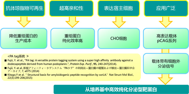
  PA tag 系统的亲和性和特异性优于现有标签系统,抗体琼脂糖可再生 60 次,极大地降低成本,可用于免疫细胞化学、免疫印迹和流式细胞术,是动物宿主细胞重组蛋白生产的有效工具。

原理

PA Tag 新型标签系统                              PA Tag

载体列表


产品名称(载体名称)

抗性

规格

产品编号

大肠杆菌抗性

真核细胞抗性

pCAG-Ble PA tag-C

Bleomycin

20 μg

161-26861

pCAG-Ble PA tag-N Signal Plus

20 μg

168-26871

pCAG-Bsd PA tag-C

Blasticidin S

20 μg

165-26881

pCAG-Bsd PA tag-N Signal Plus

20 μg

162-26891

pCAG-Hyg PA tag-C

Hygromycin B

20 μg

165-26901

pCAG-Hyg PA tag-N Signal Plus

20 μg

162-26911

pCAG-Neo PA tag-C

Kanamycin

G418

20 μg

169-26921

pCAG-Neo PA tag-N Signal Plus

20 μg

166-26931

优点、特色


PA Tag 新型标签系统                              PA Tag

案例、应用


【使用例子:PA 标签抗体,大鼠单克隆抗体(NZ-1)】


  NZ-1 是特异性识别 PA tag(GVAMPGAEDDVV)的单克隆抗体,能用于检测动物细胞表达的 PA tag 融合蛋白,亲和性很高并能简单地用肽洗脱!已确认不存在交叉反应的物种包括小鼠、大鼠、仓鼠和犬类。

PA Tag 新型标签系统                              PA Tag


浓度:       标签表示(约1.0 mg/mL)

配方:       1× PBS(pH 7.5)水溶液

克隆号:    NZ-1

亚型:       IgG2a

应用:  免疫印迹 0.01 – 1 μg/mL  

                 免疫沉淀 10 – 50 μg/assay  

                 流式细胞术 0.1 – 10 μg/mL  

                 免疫细胞化学 0.1 – 10 μg/mL

保存条件:2–10°C 避免反复冻融


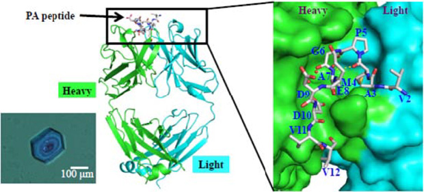
【NZ-1 Fab和PA肽的复合体结构分析】

PA Tag 新型标签系统                              PA Tag


标记抗PA tag抗体

PA tag-EGFP-6×His tag,重组,溶液


亲和标签比较

      亲和标签

     TARGET

             PA

       6 x His

       c-Mys

      HA

      GST

序列

(YPGQ)5V

GVAMPGAEDDVV

HHHHHH

EQKLISEEDL

YPYDVPDYA

残基数

21

12

6

10

9

218

分子量(kDa)

2.34

1.16

0.84

2.34

1.16

0.84

配位子

TARGET tag

PA tag

Ni, Co, Zn, Cu

c-Myc tag

HA tag

GST

抗体琼脂糖的

再利用

最高可再生循环利用40次

最高可再生循环利用60次

不可再生

不可再生

不可再生

不可再生

重组蛋白

纯化成本

+

+

+

++

++

+

单克隆抗体结合力(KD(M))(数字越小,亲和力越高)

1.0×10⁻⁸

4.9×10⁻¹⁰

Ni-NTA:

 1.0×10⁻⁵~6

2.2×10⁻⁹

2.8×10⁻⁹

Glutathione:

  1.0×10⁻⁵~6

亲和性

++++

+++++

+++

++++

++++

++++

和动物细胞外(培养基)的蛋白非特异性结合

+

+

+++++

+++

+++

++++

和动物细胞内蛋白的非特异性结合

+

+

++++

++++

+++

++++

肽洗脱

+++

+++

咪唑洗脱

++

++

谷胱甘肽洗脱

本比较表由富士胶片和光独立完成。样品、检测方法及手法不同有可能产生的特异性有变化,不能保证各种亲和标签的性能。

Target tag 系统和 Pa tag 系统同属Wako亲和标签系统,详情请联系经销商。        


 Wako新一代超亲和性的亲和标签系统“PA Tag System”http://labchem.fujifilm-wako.com.cn/resources/show/14.html


1. Fujii, Y. et al., "PA tag: A versatile protein tagging system using a super high affinity antibody against dodecapeptide

     derived from human podoplanin.", Protein Exp. Purif., 95, 240-247(2014).

2. Fujii, Y. et al. 新規アフィニティータグシステム“PAタグ”の利用法~蛋白質の精製および検出~蛋白質科学会アーカイブ, 7,e075

      (2014)

3. Kitago,Y et al.“ Structural basis for amyloidogenic peptide recognition by sorLA.”Nat Struct Mol Biol., 22(3):199-206(2015)


产品编号 产品名称 产品规格 产品等级
161-26861 pCAG-Ble PA tag-C 
 pCAG-Ble PA 标签-C
20 μg for Genetic Research
168-26871 pCAG-Ble PA tag-N Signal Plus
 pCAG-Ble PA 标签-N 信号 增加型
20 μg for Genetic Research
165-26881 pCAG-Bsd PA tag-C
 pCAG-Bsd PA 标签-C
20 μg for Genetic Research
162-26891 pCAG-Bsd PA tag-N Signal Plus
 pCAG-Bsd PA 标签-N 信号 增加型
20 μg for Genetic Research
165-26901 pCAG-Hyg PA tag-C 
pCAG-Neo PA 标签-C
2 0μg for Genetic Research
162-26911 pCAG-Hyg PA tag-N Signal Plus
 pCAG-Hyg PA 标签-N 信号 增加型
20 μg for Genetic Research
169-26921 pCAG-Neo PA tag-C
 pCAG-Neo PA 标签-C
20 μg for Genetic Research
166-26931 pCAG-Neo PA tag-N Signal Plus
 pCAG-Neo PA 标签-N 信号 增加型
20 μg for Genetic Research

Phos-tag™ 琼脂糖 Phos-tag™ Agarose

  • 产品特性
  • 相关资料
  • Q&A
  • 参考文献

Phos-tag™ AgarosePhos-tag™ 琼脂糖                              Phos-tag™ Agarose

亲和层析纯化磷酸化蛋白

  填入色谱柱中使用。可分离、纯化、浓缩磷酸化蛋白。不使用界面活性剂、还原剂,可得到状态近似生物体内的磷酸化蛋白。


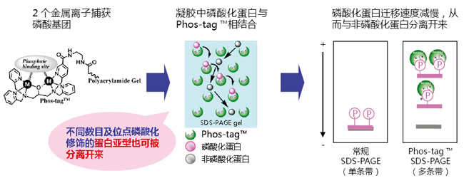
原理


Phos-tag™ 琼脂糖                              Phos-tag™ Agarose


优点、特色


 缓冲液不含有界面活性剂、还原剂。

 与亲和层析方法类似。

 可在1小时内纯化磷酸化蛋白。

 Phos-tag™ Agarose 捕获结合到 Tyr、Thr、Ser、Asp、His 等氨基酸、糖类、脂类上的无机磷酸根和大二价磷酸根

 可在生理条件下(pH7.5)捕捉蛋白。

 纯化后的产物可用于 Co-IP 实验和其他蛋白活性实验。

案例、应用:


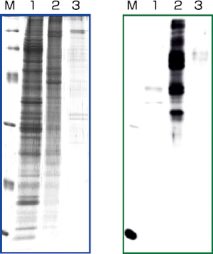
【使用例子:A431 裂解液中的磷酸化蛋白的纯化】


  把Phos-tag™ 填充到柱里,再加上 A431 裂解液。

  SYPRO Ruby 染色(左图)再使用 Anti-p Tyr 抗体进行免疫印迹(右图),检测出结果。

  结果确认磷酸化蛋白浓缩在柱吸附层里。


  M:分子量标记

  Lane 1:未吸附层

  Lane 2:吸附层

  Lane 3:柱清洗层

Phos-tag™ 琼脂糖                              Phos-tag™ Agarose

Phos-tag™ 系列

磷酸化蛋白新方法!

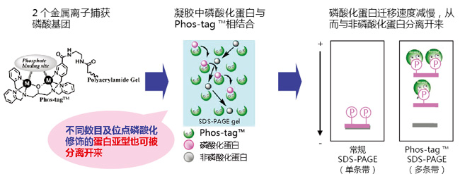
  Phos-tag™ 是一种能与磷酸离子特异性结合的功能性分子。它可用于磷酸化蛋白的分离(Phos-tag™ Acrylamide)、Western Blot 检测(Phos-tag™ Biotin)、蛋白纯化 (Phos-tag™ Agarose)及质谱分析 MALDI-TOF/MS (Phos-tag™ Mass Analytical Kit)。


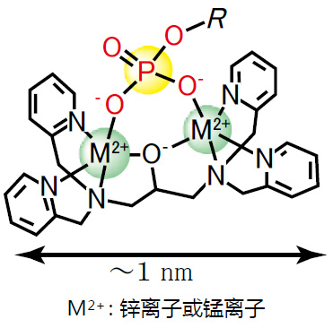
Phos-tag™ 的基本结构


Phos-tag™ 琼脂糖                              Phos-tag™ Agarose


特点


与 -2 价磷酸根离子的亲和性和选择性高于其它阴离子

在 pH 5-8 的生理环境下生成稳定的复合物

原理


Phos-tag™ 琼脂糖                              Phos-tag™ Agarose

相关应用


Phos-tag™ 琼脂糖                              Phos-tag™ Agarose

相关产品

 产品名称

 用  途

 Phos-tag™ Acrylamide

 分离SDS – PAGE 分离不同磷酸化水平的蛋白

 SuperSep Phos-tag™

 分离预制胶中含有 50 μM Phos-tag™ Acrylamide

 Phos-tag™ Biotin

 检测代替 Western Blot 检测中的磷酸化抗体

 Phos-tag™ Agarose

 纯化通用柱层析,纯化磷酸化蛋白

 Phos-tag™ Mass

 Analytical Kit

 分析:用于质谱 MALDI-TOF/MS 分析,提高磷酸化分子的检测灵敏度


phos-tag™ 由日本广岛大学研究生院医齿药学综合研究科医药分子功能科学研究室开发。

更多产品信息,请点击:http://phos-tag.jp

Phos-tag™ 琼脂糖                              Phos-tag™ Agarose

Phos-tag 第6版说明书

Phos-tag™ 琼脂糖                              Phos-tag™ Agarose

Phos-tag系列 ver. 8


Phos-tag™ 琼脂糖                              Phos-tag™ Agarose

说明书

【参考文献】


·  Conversion of graded phosphorylation into switch-like nuclear translocation via autoregulatory mechanisms in ERK signalling[J].Nature communications, 2016, 7,Shindo Y, Iwamoto K, Mouri K, et al.

·  PTEN modulates EGFR late endocytic trafficking and degradation by dephosphorylating Rab7[J]. Nature communications, 2016, 7,Shinde S R, Maddika S.

·  Feedback control of ErbB2 via ERK-mediated phosphorylation of a conserved threonine in the juxtamembrane domain[J]. Scientific Reports, 2016, 6: 31502,Kawasaki Y, Sakimura A, Park C M, et al.

·  Plastid-nucleus communication involves calcium-modulated MAPK signalling[J]. Nature Communications, 2016, 7,Guo H, Feng P, Chi W, et al.

·  Sequential domain assembly of ribosomal protein S3 drives 40S subunit maturation[J]. Nature communications, 2016, 7,Mitterer V, Murat G, Réty S, et al.

·  Phos-tag analysis of Rab10 phosphorylation by LRRK2: a powerful assay for assessing kinase function and inhibitors[J]. Biochemical Journal, 2016: BCJ20160557,Ito G, Katsemonova K, Tonelli F, et al.

·  Analysis of phosphorylation of the myosin targeting subunit of smooth muscle myosin light chain phosphatase by Phos-tag SDS-PAGE[J]. The FASEB Journal, 2016, 30(1 Supplement): 1209.1-1209.1,Walsh M P, MacDonald J A, Sutherland C.

·  Using Phos-Tag in Western Blotting Analysis to Evaluate Protein Phosphorylation[J]. Kidney Research: Experimental Protocols, 2016: 267-277,Horinouchi T, Terada K, Higashi T, et al.

·  The Abundance of Nonphosphorylated Tau in Mouse and Human Tauopathy Brains Revealed by the Use of Phos-Tag Method[J]. The American journal of pathology, 2016, 186(2): 398-409,Kimura T, Hatsuta H, Masuda-Suzukake M, et al.

·  Phos-tag SDS-PAGE resolves agonist-and isoform-specific activation patterns for PKD2 and PKD3 in cardiomyocytes and cardiac fibroblasts[J]. Journal of Molecular and Cellular Cardiology, 2016,Qiu W, Steinberg S F.

·  Analysis of phosphorylation of the myosin-targeting subunit of myosin light chain phosphatase by Phos-tag SDS-PAGE[J]. American Journal of Physiology-Cell Physiology, 2016, 310(8): C681-C691,Sutherland C, MacDonald J A, Walsh M P.

·  Electrochemical biosensor for protein kinase A activity assay based on gold nanoparticles-carbon nanospheres, phos-tag-biotin and β-galactosidase[J]. Biosensors and Bioelectronics, 2016, 86: 508-515,Zhou Y, Yin H, Li X, et al.

·  Validation of Cis and Trans Modes in Multistep Phosphotransfer Signaling of Bacterial Tripartite Sensor Kinases by Using Phos-Tag SDS-PAGE[J]. PloS one, 2016, 11(2): e0148294,Kinoshita-Kikuta E, Kinoshita E, Eguchi Y, et al.

·  Phosphopeptide Detection with Biotin-Labeled Phos-tag[J]. Phospho-Proteomics: Methods and Protocols, 2016: 17-29,Kinoshita-Kikuta E, Kinoshita E, Koike T.

·  A Phos‐tag SDS‐PAGE method that effectively uses phosphoproteomic data for profiling the phosphorylation dynamics of MEK1[J]. Proteomics, 2016,Kinoshita E, Kinoshita‐Kikuta E, Kubota Y, et al.

·  Difference gel electrophoresis of phosphoproteome: U.S. Patent Application 15/004,339[P]. 2016-1-22,Tao W A, Wang L.

·  ERK1/2-induced phosphorylation of R-Ras GTPases stimulates their oncogenic potential[J]. Oncogene, 2016,Frémin C, Guégan J P, Plutoni C, et al.

·  Microtubules Inhibit E-Cadherin Adhesive Activity by Maintaining Phosphorylated p120-Catenin in a Colon Carcinoma Cell Model[J]. PloS one, 2016, 11(2): e0148574,Maiden S L, Petrova Y I, Gumbiner B M.

·  Serine 231 and 257 of Agamous-like 15 are phosphorylated in floral receptacles[J]. Plant Signaling & Behavior, 2016, 11(7): e1199314,Patharkar O R, Macken T A, Walker J C.

·  A small molecule pyrazolo [3, 4-d] pyrimidinone inhibitor of zipper-interacting protein kinase suppresses calcium sensitization of vascular smooth muscle[J]. Molecular pharmacology, 2016, 89(1): 105-117,MacDonald J A, Sutherland C, Carlson D A, et al.

·  The RNA polymerase II C-terminal domain phosphatase-like protein FIERY2/CPL1 interacts with eIF4AIII and is essential for nonsense-mediated mRNA decay in Arabidopsis[J]. The Plant Cell, 2016: TPC2015-00771-RA,Chen T, Qin T, Ding F, et al.

·  Vasorelaxant Effect of 5′-Methylthioadenosine Obtained from Candida utilis Yeast Extract through the Suppression of Intracellular Ca2+ Concentration in Isolated Rat Aorta[J]. Journal of agricultural and food chemistry, 2016, 64(17): 3362-3370,Kumrungsee T, Akiyama S, Saiki T, et al.

·  Inhibition of deubiquitinating activity of USP14 decreases tyrosine hydroxylase phosphorylated at Ser19 in PC12D cells[J]. Biochemical and biophysical research communications, 2016, 472(4): 598-602,Nakashima A, Ohnuma S, Kodani Y, et al.

·  Actin Tyrosine-53-Phosphorylation in Neuronal Maturation and Synaptic Plasticity[J]. The Journal of Neuroscience, 2016, 36(19): 5299-5313,Bertling E, Englund J, Minkeviciene R, et al.

·  AMPK-dependent phosphorylation of lipid droplet protein PLIN2 triggers its degradation by CMA[J]. Autophagy, 2016, 12(2): 432-438,Kaushik S, Cuervo A M.

·  Myocardin-related transcription factor a and yes-associated protein exert dual control in G protein-coupled receptor-and RhoA-mediated transcriptional regulation and cell proliferation[J]. Molecular and cellular biology, 2016, 36(1): 39-49,Olivia M Y, Miyamoto S, Brown J H.

·  Extensive phosphorylation of AMPA receptors in neurons[J]. Proceedings of the National Academy of Sciences, 2016, 113(33): E4920-E4927,Diering G H, Heo S, Hussain N K, et al.

·  The transmembrane region of guard cell SLAC1 channels perceives CO2 signals via an ABA-independent pathway in Arabidopsis[J]. The Plant Cell, 2016, 28(2): 557-567,Yamamoto Y, Negi J, Wang C, et al.

·  The Hippo pathway mediates inhibition of vascular smooth muscle cell proliferation by cAMP[J]. Journal of molecular and cellular cardiology, 2016, 90: 1-10,Kimura T E, Duggirala A, Smith M C, et al.

·  Atg13 is essential for autophagy and cardiac development in mice[J]. Molecular and cellular biology, 2016, 36(4): 585-595,Kaizuka T, Mizushima N.

·  The ChrSA and HrrSA two-component systems are required for transcriptional regulation of the hemA promoter in Corynebacterium diphtheriae[J]. Journal of Bacteriology, 2016: JB. 00339-16,Burgos J M, Schmitt M P.

·  Intergenic Variable-Number Tandem-Repeat Polymorphism Upstream of rocA Alters Toxin Production and Enhances Virulence in Streptococcus pyogenes[J]. Infection and Immunity, 2016, 84(7): 2086-2093,Zhu L, Olsen R J, Horstmann N, et al.

·  Receptor for advanced glycation end products (RAGE) knockout reduces fetal dysmorphogenesis in murine diabetic pregnancy[J]. Reproductive Toxicology, 2016, 62: 62-70,Ejdesjö A, Brings S, Fleming T, et al.

·  Aurora kinase-induced phosphorylation excludes transcription factor RUNX from the chromatin to facilitate proper mitotic progression[J]. Proceedings of the National Academy of Sciences, 2016, 113(23): 6490-6495,Chuang L S H, Khor J M, Lai S K, et al.

·  Quantitative phosphoproteomics of protein kinase SnRK1 regulated protein phosphorylation in Arabidopsis under submergence[J]. Journal of experimental botany, 2016: erw107,Cho H Y, Wen T N, Wang Y T, et al.

·  Temporal regulation of lipin activity diverged to account for differences in mitotic programs[J]. Current Biology, 2016, 26(2): 237-243,Makarova M, Gu Y, Chen J S, et al.

·  Block of CDK1‐dependent polyadenosine elongation of Cyclin B mRNA in metaphase‐i‐arrested starfish oocytes is released by intracellular pH elevation upon spawning[J]. Molecular reproduction and development, 2016, 83(1): 79-87,Ochi H, Aoto S, Tachibana K, et al.

·  Mitotic Exit Function of Polo-like Kinase Cdc5 Is Dependent on Sequential Activation by Cdk1[J]. Cell reports, 2016, 15(9): 2050-2062,Rodriguez-Rodriguez J A, Moyano Y, Játiva S, et al.

·  PLK2 phosphorylates and inhibits enriched TAp73 in human osteosarcoma cells[J]. Cancer medicine, 2016, 5(1): 74-87,Hu Z B, Liao X H, Xu Z Y, et al.

·  Phosphorylated TDP-43 becomes resistant to cleavage by calpain: A regulatory role for phosphorylation in TDP-43 pathology of ALS/FTLD[J]. Neuroscience research, 2016, 107: 63-69,Yamashita T, Teramoto S, Kwak S.

·  The Pch2 AAA+ ATPase promotes phosphorylation of the Hop1 meiotic checkpoint adaptor in response to synaptonemal complex defects[J]. Nucleic acids research, 2016: gkw506,Herruzo E, Ontoso D, González-Arranz S, et al.

·  An optimized guanidination method for large‐scale proteomic studies[J]. Proteomics, 2016,Ye J, Zhang Y, Huang L, et al.

·  Expression and purification of the kinase domain of PINK1 in Pichia pastoris[J]. Protein Expression and Purification, 2016,Wu D, Qu L, Fu Y, et al.

·  BRI2 and BRI3 are functionally distinct phosphoproteins[J]. Cellular signalling, 2016, 28(1): 130-144,Martins F, Rebelo S, Santos M, et al.

·  Identification of glycoproteins associated with HIV latently infected cells using quantitative glycoproteomics[J]. Proteomics, 2016,Yang W, Jackson B, Zhang H.

·  Regulation of Beclin 1 Protein Phosphorylation and Autophagy by Protein Phosphatase 2A (PP2A) and Death-associated Protein Kinase 3 (DAPK3)[J]. Journal of Biological Chemistry, 2016, 291(20): 10858-10866,Fujiwara N, Usui T, Ohama T, et al.

·  Regulatory Implications of Structural Changes in Tyr201 of the Oxygen Sensor Protein FixL[J]. Biochemistry, 2016, 55(29): 4027-4035,Yamawaki T, Ishikawa H, Mizuno M, et al.

·  Histone demethylase Jmjd3 regulates osteoblast apoptosis through targeting anti-apoptotic protein Bcl-2 and pro-apoptotic protein Bim[J]. Biochimica et Biophysica Acta (BBA)-Molecular Cell Research, 2016, 1863(4): 650-659,Yang D, Okamura H, Teramachi J, et al.

·  Analysis of Molecular Species Profiles of Ceramide-1-phosphate and Sphingomyelin Using MALDI-TOF Mass Spectrometry[J]. Lipids, 2016, 51(2): 263-270,Yamashita R, Tabata Y, Iga E, et al.

·  Highly sensitive myosin phosphorylation analysis in the renal afferent arteriole[J]. Journal of Smooth Muscle Research, 2016, 52(0): 45-55,Takeya K.

·  Functional dissection of the CroRS two-component system required for resistance to cell wall stressors in Enterococcus faecalis[J]. Journal of bacteriology, 2016, 198(8): 1326-1336,Kellogg S L, Kristich C J.

·  Regulation of mitogen-activated protein kinase by protein kinase C and mitogen-activated protein kinase phosphatase-1 in vascular smooth muscle[J]. American Journal of Physiology-Cell Physiology, 2016, 310(11): C921-C930,Trappanese D M, Sivilich S, Ets H K, et al.

·  ModProt: a database for integrating laboratory and literature data about protein post-translational modifications[J]. Journal of Electrophoresis, 2016, 60(1): 1-4,Kimura Y, Toda T, Hirano H.

·  The C-ETS2-TFEB Axis Promotes Neuron Survival under Oxidative Stress by Regulating Lysosome Activity[J]. Oxidative medicine and cellular longevity, 2016,Ma S, Fang Z, Luo W, et al.

·  Essential role of the PSI–LHCII supercomplex in photosystem acclimation to light and/or heat conditions by state transitions[J]. Photosynthesis Research, 2016: 1-10,Marutani Y, Yamauchi Y, Higashiyama M, et al.

·  Identification of a redox-modulatory interaction between selenoprotein W and 14-3-3 protein[J]. Biochimica et Biophysica Acta (BBA)-Molecular Cell Research, 2016, 1863(1): 10-18,Jeon Y H, Ko K Y, Lee J H, et al.

·  Effects of hydrogen sulfide on the heme coordination structure and catalytic activity of the globin-coupled oxygen sensor AfGcHK[J]. BioMetals, 2016, 29(4): 715-729,Fojtikova V, Bartosova M, Man P, et al.

·  Identification and functional analysis of phosphorylation in Newcastle disease virus phosphoprotein[J]. Archives of virology, 2016: 1-14,Qiu X, Zhan Y, Meng C, et al.

·  Increased level of phosphorylated desmin and its degradation products in heart failure[J]. Biochemistry and Biophysics Reports, 2016, 6: 54-62,Bouvet M, Dubois-Deruy E, Alayi T D, et al.

·  Profiling DNA damage-induced phosphorylation in budding yeast reveals diverse signaling networks[J]. Proceedings of the National Academy of Sciences, 2016: 201602827,Zhou C, Elia A E H, Naylor M L, et al.

·  Unexpected properties of sRNA promoters allow feedback control via regulation of a two-component system[J]. Nucleic Acids Research, 2016: gkw642,Brosse A, Korobeinikova A, Gottesman S, et al.

·  Evolution of ZnII–Macrocyclic Polyamines to Biological Probes and Supramolecular Assembly[J]. Macrocyclic and Supramolecular Chemistry: How Izatt-Christensen Award Winners Shaped the Field, 2016: 415,Kimura E, Koike T, Aoki S.

·  Phosphopeptide Enrichment Using Various Magnetic Nanocomposites: An Overview[J]. Phospho-Proteomics: Methods and Protocols, 2016: 193-209,Batalha Í L, Roque A C A.

·  In vivo phosphorylation of a peptide tag for protein purification[J]. Biotechnology letters, 2016, 38(5): 767-772,Goux M, Fateh A, Defontaine A, et al.

·  Regulation of cell reversal frequency in Myxococcus xanthus requires the balanced activity of CheY‐like domains in FrzE and FrzZ[J]. Molecular microbiology, 2016,Kaimer C, Zusman D R.

·  Elevation of cortical serotonin transporter activity upon peripheral immune challenge is regulated independently of p38 mitogen‐activated protein kinase activation and transporter phosphorylation[J]. Journal of neurochemistry, 2016, 137(3): 423-435,Schwamborn R, Brown E, Haase J.

·  The Yeast Cyclin-Dependent Kinase Routes Carbon Fluxes to Fuel Cell Cycle Progression[J]. Molecular cell, 2016, 62(4): 532-545,Ewald J C, Kuehne A, Zamboni N, et al.

·  Two Degradation Pathways of the p35 Cdk5 (Cyclin-dependent Kinase) Activation Subunit, Dependent and Independent of Ubiquitination[J]. Journal of Biological Chemistry, 2016, 291(9): 4649-4657,Takasugi T, Minegishi S, Asada A, et al.

·  Increased level of phosphorylated desmin and its degradation products in heart failure[J]. Biochemistry and Biophysics Reports. 2016,Bouvet M, Dubois-Deruy E, Alayi T D, et al.

·  a high‐affinity LCO‐binding protein of Medicago truncatula, interacts with LYK3, a key symbiotic receptor[J]. FEBS letters, 2016, 590(10): 1477-1487,Fliegmann J, Jauneau A, Pichereaux C, et al. LYR3,

·  Nek1 Regulates Rad54 to Orchestrate Homologous Recombination and Replication Fork Stability[J]. Molecular Cell, 2016,Spies J, Waizenegger A, Barton O, et al.

·  PhostagTM-gel retardation and in situ thylakoid kinase assay for determination of chloroplast protein phosphorylation targets[J]. Endocytobiosis and Cell Research, 2016, 27(2): 62-70,Dytyuk Y, Flügge F, Czarnecki O, et al.

·  Luteinizing Hormone Causes Phosphorylation and Activation of the cGMP Phosphodiesterase PDE5 in Rat Ovarian Follicles, Contributing, Together with PDE1 Activity, to the Resumption of Meiosis[J]. Biology of reproduction, 2016: biolreprod. 115.135897,Egbert J R, Uliasz T F, Shuhaibar L C, et al.

·  Newby, AC, & Bond, M.(2016). The Hippo pathway mediates inhibition of vascular smooth muscle cell proliferation by cAMP[J]. Journal of Molecular and Cellular Cardiology, 2016, 90: 1-10,Kimura-Wozniak T, Duggirala A, Smith M C, et al. G.

·  Yeast lacking the amphiphysin family protein Rvs167 is sensitive to disruptions in sphingolipid levels[J]. The FEBS Journal, 2016, 283(15): 2911-2928,Toume M, Tani M.

·  Regulation of CsrB/C sRNA decay by EIIAGlc of the phosphoenolpyruvate: carbohydrate phosphotransferase system[J]. Molecular microbiology, 2016, 99(4): 627-639,Leng Y, Vakulskas C A, Zere T R, et al.

·  The Late S-Phase Transcription Factor Hcm1 Is Regulated through Phosphorylation by the Cell Wall Integrity Checkpoint[J]. Molecular and cellular biology, 2016: MCB. 00952-15,Negishi T, Veis J, Hollenstein D, et al.

·  Validation of chemical compound library screening for transcriptional co‐activator with PDZ‐binding motif inhibitors using GFP‐fused transcriptional co‐activator with PDZ‐binding motif[J]. Cancer science, 2016, 107(6): 791-802,Nagashima S, Maruyama J, Kawano S, et al.

·  ULK1/2 Constitute a Bifurcate Node Controlling Glucose Metabolic Fluxes in Addition to Autophagy[J]. Molecular cell, 2016, 62(3): 359-370,Li T Y, Sun Y, Liang Y, et al.

·  Spatiotemporal dynamics of Oct4 protein localization during preimplantation development in mice[J]. Reproduction, 2016: REP-16-0277,Fukuda A, Mitani A, Miyashita T, et al.

·  The tandemly repeated NTPase (NTPDase) from Neospora caninum is a canonical dense granule protein whose RNA expression, protein secretion and phosphorylation coincides with the tachyzoite egress[J]. Parasites & Vectors, 2016, 9(1): 1,Pastor-Fernández I, Regidor-Cerrillo J, Álvarez-García G, et al.

·  Interaction Analysis of a Two-Component System Using Nanodiscs[J]. PloS one, 2016, 11(2): e0149187,Hörnschemeyer P, Liss V, Heermann R, et al.

·  Constitutive Activation of PINK1 Protein Leads to Proteasome-mediated and Non-apoptotic Cell Death Independently of Mitochondrial Autophagy[J]. Journal of Biological Chemistry, 2016, 291(31): 16162-16174,Akabane S, Matsuzaki K, Yamashita S, et al.

·  p38β Mitogen-Activated Protein Kinase Modulates Its Own Basal Activity by Autophosphorylation of the Activating Residue Thr180 and the Inhibitory Residues Thr241 and Ser261[J]. Molecular and cellular biology, 2016, 36(10): 1540-1554,Beenstock J, Melamed D, Mooshayef N, et al.

·  Lysophosphatidylcholine acyltransferase 1 protects against cytotoxicity induced by polyunsaturated fatty acids[J]. The FASEB Journal, 2016, 30(5): 2027-2039,Akagi S, Kono N, Ariyama H, et al.

·  Characterization of a herpes simplex virus 1 (HSV-1) chimera in which the Us3 protein kinase gene is replaced with the HSV-2 Us3 gene[J]. Journal of virology, 2016, 90(1): 457-473,Shindo K, Kato A, Koyanagi N, et al.

·  Generation of phospho‐ubiquitin variants by orthogonal translation reveals codon skipping[J]. FEBS letters, 2016, 590(10): 1530-1542,George S, Aguirre J D, Spratt D E, et al.

·  Evolution of KaiC-Dependent Timekeepers: A Proto-circadian Timing Mechanism Confers Adaptive Fitness in the Purple Bacterium Rhodopseudomonas palustris[J]. PLoS Genet, 2016, 12(3): e1005922,Ma P, Mori T, Zhao C, et al.

·  Phosphorylation of Bni4 by MAP kinases contributes to septum assembly during yeast cytokinesis[J]. FEMS Yeast Research, 2016, 16(6): fow060,Pérez J, Arcones I, Gómez A, et al.

·  Alteration of Antiviral Signalling by Single Nucleotide Polymorphisms (SNPs) of Mitochondrial Antiviral Signalling Protein (MAVS)[J]. PloS one, 2016, 11(3): e0151173,Xing F, Matsumiya T, Hayakari R, et al.

·  Arm-in-arm response regulator dimers promote intermolecular signal transduction[J]. Journal of bacteriology, 2016, 198(8): 1218-1229,Baker A W, Satyshur K A, Morales N M, et al.

·  The lsh/ddm1 homolog mus-30 is required for genome stability, but not for dna methylation in neurospora crassa[J]. PLoS Genet, 2016, 12(1): e1005790,Basenko E Y, Kamei M, Ji L, et al.

·  Fine tuning chloroplast movements through physical interactions between phototropins[J]. Journal of Experimental Botany, 2016: erw265,Sztatelman O, Łabuz J, Hermanowicz P, et al.

·  Characterization of the Neospora caninum NcROP40 and NcROP2Fam-1 rhoptry proteins during the tachyzoite lytic cycle[J]. Parasitology, 2016, 143(01): 97-113,Pastor-Fernandez I, Regidor-Cerrillo J, Jimenez-Ruiz E, et al.

·  Transcriptional Profile during Deoxycholate-Induced Sporulation in a Clostridium perfringens Isolate Causing Foodborne Illness[J]. Applied and environmental microbiology, 2016, 82(10): 2929-2942,Yasugi M, Okuzaki D, Kuwana R, et al.

·  Timely Closure of the Prospore Membrane Requires SPS1 and SPO77 in Saccharomyces cerevisiae[J]. Genetics, 2016: genetics. 115.183939,Paulissen S M, Slubowski C J, Roesner J M, et al.

·  DDK dependent regulation of TOP2A at centromeres revealed by a chemical genetics approach[J]. Nucleic Acids Research, 2016: gkw626,Wu K Z L, Wang G N, Fitzgerald J, et al.

·  OVATE Family Protein 8 Positively Mediates Brassinosteroid Signaling through Interacting with the GSK3-like Kinase in Rice[J]. PLoS Genet, 2016, 12(6): e1006118,Yang C, Shen W, He Y, et al.

·  Epithelial Sel1L is required for the maintenance of intestinal homeostasis[J]. Molecular biology of the cell, 2016, 27(3): 483-490, Sun S, Lourie R, Cohen S B, et al.

·  Effect of Sodium Dodecyl Sulfate Concentration on Supramolecular Gel Electrophoresis[J]. ChemNanoMat, 2016,Tazawa S, Kobayashi K, Yamanaka M.

·  Intergenic VNTR Polymorphism Upstream of rocA Alters Toxin Production and Enhances Virulence in Streptococcus pyogenes[J]. Infection and immunity, 2016: IAI. 00258-16,Zhu L, Olsen R J, Horstmann N, et al.

·  Ajuba Phosphorylation by CDK1 Promotes Cell Proliferation and Tumorigenesis[J]. Journal of Biological Chemistry, 2016: jbc. M116. 722751,Chen X, Stauffer S, Chen Y, et al.

·  Editorial: International Plant Proteomics Organization (INPPO) World Congress 2014[J]. Frontiers in Plant Science, 2016, 7,Heazlewood J L, Jorrín-Novo J V, Agrawal G K, et al.

·  Phosphoinositide kinase signaling controls ER-PM cross-talk[J]. Molecular biology of the cell, 2016, 27(7): 1170-1180,Omnus D J, Manford A G, Bader J M, et al.

·  A multiple covalent crosslinked soft hydrogel for bioseparation[J]. Chemical Communications, 2016, 52(15): 3247-3250,Liu Z, Fan L, Xiao H, et al.

·  Advances in crop proteomics: PTMs of proteins under abiotic stress[J]. Proteomics, 2016, 16(5): 847-865,Wu X, Gong F, Cao D, et al.

·  Cyclin-Dependent Kinase Co-Ordinates Carbohydrate Metabolism and Cell Cycle in S. cerevisiae[J]. Molecular cell, 2016, 62(4): 546-557,Zhao G, Chen Y, Carey L, et al.

·  Carbon Monoxide Gas Is Not Inert, but Global, in Its Consequences for Bacterial Gene Expression, Iron Acquisition, and Antibiotic Resistance[J]. Antioxidants & redox signaling, 2016,Wareham L K, Begg R, Jesse H E, et al.

·  Two-layer regulation of PAQR3 on ATG14-linked class III PtdIns3K activation upon glucose starvation[J]. Autophagy, 2016: 1-2,Xu D, Wang Z, Chen Y.

·  Regulation of sphingolipid biosynthesis by the morphogenesis checkpoint kinase Swe1[J]. Journal of Biological Chemistry, 2016, 291(5): 2524-2534,Chauhan N, Han G, Somashekarappa N, et al.

·  PAX5 tyrosine phosphorylation by SYK co-operatively functions with its serine phosphorylation to cancel the PAX5-dependent repression of BLIMP1: A mechanism for antigen-triggered plasma cell differentiation[J]. Biochemical and biophysical research communications, 2016, 475(2): 176-181,Inagaki Y, Hayakawa F, Hirano D, et al.

·  A Combined Computational and Genetic Approach Uncovers Network Interactions of the Cyanobacterial Circadian Clock[J]. Journal of Bacteriology, 2016: JB. 00235-16,Boyd J S, Cheng R R, Paddock M L, et al.

·  HuR mediates motility of human bone marrow-derived mesenchymal stem cells triggered by sphingosine 1-phosphate in liver fibrosis[J]. Journal of Molecular Medicine, 2016: 1-14,Chang N, Ge J, Xiu L, et al.

·  Combined replacement effects of human modified β-hexosaminidase B and GM2 activator protein on GM2 gangliosidoses fibroblasts[J]. Biochemistry and Biophysics Reports, 2016,Kitakaze K, Tasaki C, Tajima Y, et al.

·  Roseotoxin B Improves Allergic Contact Dermatitis through a Unique Anti-inflammatory Mechanism Involving Excessive Activation of Autophagy in Activated T-Lymphocytes[J]. Journal of Investigative Dermatology, 2016,Wang X, Hu C, Wu X, et al.

References on Phos-tag™ Chemistry

Matrix-assisted laser desorption/ionization time-of-flight mass spectrometry of phosphorylated compounds using a novel phosphate capture moleculeRapid Communications of Mass Spectrometry17, 2075-2081 (2003), H. Takeda, A. Kawasaki, M. Takahashi, A. Yamada, and T. Koike 

Recognition of phosphate monoester dianion by an alkoxide-bridged dinuclear zinc (II) complexDalton Transactions, 1189-1193 (2004), E. Kinoshita, M. Takahashi, H. Takeda, M. Shiro, and T. Koike

Quantitative analysis of lysophosphatidic acid by time-of-flight mass spectrometry using a phosphate capture molecule, Journal of Lipid Research45, 2145-2150 (2004), T. Tanaka, H. Tsutsui, K. Hirano, T. Koike, A. Tokumura, and K. Satouchi

Production of 1,2-Didocosahexaenoyl Phosphatidylcholine by Bonito Muscle Lysophosphatidylcholine/TransacylaseJournal of Biochemistry,136, 477-483 (2004), K. Hirano, H. Matsui, T. Tanaka, F. Matsuura, K. Satouchi, and T. Koike

Novel immobilized zinc(II) affinity chromatography for phosphopeptides and phosphorylated proteins, Journal of Separation Science, 28, 155-162 (2005), E. Kinoshita, A. Yamada, H. Takeda, E. Kinoshita-Kikuta, and T. Koike

Detection and Quantification of On-Chip Phosphorylated Peptides by Surface Plasmon Resonance Imaging Techniques Using a Phosphate Capture MoleculeAnalytical Chemistry77, 3979-3985 (2005), K. Inamori, M. Kyo, Y. Nishiya, Y. Inoue, T. Sonoda, E. Kinoshita, T. Koike, and Y. Katayama

Phosphate-binding tag: A new tool to visualize phosphorylated proteins, Molecular & Cellular Proteomics, 5, 749-757 (2006), E. Kinoshita, E. Kinoshita-Kikuta, K. Takiyama, and T. Koike

Enrichment of phosphorylated proteins from cell lysate using phosphate-affinity chromatography at physiological pHProteomics, 6, 5088-5095 (2006), E. Kinoshita-Kikuta, E. Kinoshita, A. Yamada, M. Endo, and T. Koike

Separation of a phosphorylated histidine protein using phosphate affinity polyacrylamide gel electrophoresis, Analytical Biochemistry360, 160-162 (2007), S. Yamada, H. Nakamura, E. Kinoshita, E. Kinoshita-Kikuta, T. Koike, and Y. Shiro

Label-free kinase profiling using phosphate-affinity polyacrylamide gel electrophresisMolecular & Cellular Proteomics, 6, 356-366 (2007), E. Kinoshita-Kikuta, Y. Aoki, E. Kinoshita, and T. Koike

A SNP genotyping method using phosphate-affinity polyacrylamide gel electrophoresis, Analytical Biochemistry361, 294-298 (2007), E. Kinoshita, E. Kinoshita-Kikuta, and T. Koike (The phosphate group at DNA-terminal is efficiently captured by Zn2+.Phos-tag.)

Identification on Membrane and Characterization of Phosphoproteins Using an Alkoxide-Bridged Dinuclear Metal Complex as a Phosphate-Binding Tag MoleculeJournal of Biomolecular Techniques18, 278-286 (2007), T. Nakanishi, E. Ando, M. Furuta, E. Kinoshita, E. Kikuta-Kinoshita, T. Koike, S. Tsunasawa, and O. Nishimura

A mobility shift detection method for DNA methylation analysis using phosphate affinity polyacrylamide gel electrophoresisAnalytical Biochemistry378, 102-104 (2008), E. Kinoshita-Kikuta, E. Kinoshita, and T. Koike

Separation of phosphoprotein isotypes having the same number of phosphate groups using phosphate- affinity SDS-PAGEProteomics, 8, 2994-3003 (2008), E. Kinoshita, E. Kinoshita-Kikuta, M. Matsubara, S. Yamada, H. Nakamura, Y. Shiro, Y. Aoki, K. Okita, and T. Koike

FANCI phosphorylation functions as a molecular switch to turn on the Fanconi anemia pathwayNature Structural & Molecular Biology15, 1138-1146 (2008), M. Ishiai, H. Kitao, A. Smogorzewska, J. Tomida, A. Kinomura, E. Uchida, A. Saberi, E. Kinoshita, E. Kinoshita-Kikuta, T. Koike, S. Tashiro, S. J. Elledge, and M. Takata

Two-dimensional phosphate affinity gel electrophoresis for the analysis of phosphoprotein isotypes Electrophoresis30, 550-559 (2009), E. Kinoshita, E. Kinoshita-Kikuta, M. Matsubara, Y. Aoki, S. Ohie, Y. Mouri, and T. Koike

Formation of lysophosphatidic acid, a wound-healing lipid, during digestion of cabbage leavesBioscience, Biotechnology, and Biochemistry,73, 1293-300 (2009), T. Tanaka, G. Horiuchi, M. Matsuoka, K. Hirano, A. Tokumura, T. Koike, and K. Satouchi

A Phos-tag-based fluorescence resonance energy transfer system for the analysis of the dephosphorylation of phosphopeptidesAnalytical Biochemistry388, 235-241, (2009), K. Takiyama, E. Kinoshita, E. Kinoshita-Kikuta, Y. Fujioka, Y. Kubo, and T. Koike

Phos-tag beads as an immunoblotting enhancer for selective detection of phosphoproteins in cell lysatesAnalytical Biochemistry389, 83-85, (2009), E. Kinoshita-Kikuta, E. Kinoshita, and T. Koike

Mobility shift detection of phosphorylation on large proteins using a Phos-tag SDS-PAGE gel strengthened with agaroseProteomics9, 4098- 4101 (2009), E. Kinoshita, E. Kinoshita-Kikuta, H. Ujihara, and T. Koike

Separation and detection of large phosphoproteins using Phos-tag SDS-PAGENature Protocols4, 1513-1521 (2009), E. Kinoshita, E. Kinoshita-Kikuta, and T. Koike

A clean-up technology for the simultaneous determination of lysophosphatidic acid and sphingosine-1-phosphate by matrix-assisted laser desorption/ionization time-of-flight mass spectrometry using a phosphate-capture molecule, Phos-tagRapid Communications in Mass Spectrometry24, 1075-1084 (2010), J. Morishige, M. Urikura, H. Takagi, K. Hirano, T. Koike, T. Tanaka, and K. Satouchi

Genotyping and mapping assay of single-nucleotide polymorphisms in CYP3A5 using DNA-binding zinc(II) complexesClinical Biochemistry43, 302-306 (2010), E. Kinoshita, E. Kinoshita-Kikuta, H. Nakashima, and T. Koike

The DNA-binding activity of mouse DNA methyltransferase 1 is ragulated phosphorylation with casein kinase 1σ/εBiochemical Journal427, 489-497 (2010), Y. Sugiyama, N. Hatano, N. Sueyoshi, I. Suetake, S. Tajima, E. Kinoshita, E. Kinoshita-Kikuta, T. Koike, and I. Kameshita


产品编号 产品名称 产品规格 产品等级
308-93563 Phos-tag™ Agarose
 Phos-tag 琼脂糖
3 mL

重组无细胞蛋白合成系统 PUREfrex® 2.0

  • 产品特性
  • 相关资料
  • Q&A
  • 参考文献

重组合无细胞蛋白合成系统重组无细胞蛋白合成系统                              PUREfrex® 2.0

PUREfrex® 2.0


◆简介


  PUREfrex® 试剂盒是在东京大学的Takuya Ueda教授所发明的PUREsystem技术基础上,新开发的一款重组合无细胞蛋白合成试剂盒。

  反应系统由蛋白质、核糖体、氨基酸和NTPs组成[1,2],其中蛋白行使转录、翻译和能量供应的功能。蛋白与核糖体为分别单独高度纯化后,再重新组合成蛋白合成系统,而非直接从大肠杆菌S30中提取。当合成蛋白时,仅需将编码目的蛋白的模板DNA或mRNA添加到反应混合液中并孵育数小时,即可完成反应。本系统的突出特色是在体外以转录相关因子重新组合一套表达系统,并可根据需要来调整反应混合物的成分,而不必担心高背景会影响下游的应用。进行蛋白表达仅需将编码目标蛋白的模板DNA或mRNA加入到反应体系中,然后孵育2-4小时即可完成反应。PUREfrex® 试剂盒的所有蛋白组分均不带标签,因此目的蛋白可融合任意标签进行纯化和检测。


重组无细胞蛋白合成系统                              PUREfrex® 2.0


>>>无细胞表达的优势<<<


● 无需制备克隆

● 无需考虑培养条件

● 无需考虑表达所需的诱导条件

● 来源于宿主的污染少


◆PUREfrex® 系列


● PUREfrex® 1.0 第一代产品    

● PUREfrex® 2.0 第二代产品,表达量更高,污染水平更低;

● RNA酶与β-半乳糖苷酶污染大大降低;

● 每1 µL反应混合物中的脂多糖(LPS)低于0.1 EU。

● PUREfrex® 2.1 更适合二硫键的形成


☆升级至PUREfrex® 2.0


1.合成原核和真核蛋白的结果显示,用PUREfrex® 2.0合成时,各种蛋白的合成量增加。


重组无细胞蛋白合成系统                              PUREfrex® 2.0

2.GFP蛋白合成的结果显示,用PUREfrex® 2.0合成时,可以观察到荧光强度增强了10倍以上(每单位反应产物)。


重组无细胞蛋白合成系统                              PUREfrex® 2.0

3.合成需要形成二硫键(SS键)的大肠杆菌酸性磷酸酶(AppA1)时,在PUREfrex® 2.0基础上,添加了DS supplement的结果显示,存在

3.氧化剂和二硫键异构酶时,活性蛋白合成量增加。


重组无细胞蛋白合成系统                              PUREfrex® 2.0


1:AppA有5个二硫键,是其中一个位点在不连续的半胱氨酸之间存在的二硫键。


4.正确高级结构蛋白的合成结果显示,存在分子伴侣2的情况下用PUREfrex® 2.0,蛋白合成量增加。


重组无细胞蛋白合成系统                              PUREfrex® 2.0


2 DnaK Mix:DnaK / DnaJ / GrpE mixture为配套分子伴侣


◆特点


● 可以同时加入多种模板进行反应,以合成Fab(带二硫键)及多聚体等带二级结构的多肽

● 可合成活细胞难以合成的强毒性蛋白

● 可直接使用PCR产物来作为模板DNA

● 单位体积内合成的蛋白量几乎恒定,不随反应体积变化而产生显著差异

● 操作简便,仅需在37℃孵育数小时

● 可以合成带标签的蛋白用于下游纯化和检测

● 产品经优化升级,合成量大大提高


◆应用


制备目的蛋白


● 原核蛋白

● 真核蛋白

● 膜蛋白

● 二硫键蛋白

● 含有非天然氨基酸的蛋白质等


蛋白基础研究


● 翻译

● 蛋白合成后折叠


体外展示技术


● 核糖体展示技术

● mRNA展示技术


◆应用实例


利用PUREfrex® 系统合成并一步纯化DHFR-His


重组无细胞蛋白合成系统                              PUREfrex® 2.0


3:模板DNA的构建方法请见"相关资料"栏或点击这里


◆添加剂(用于需要形成二硫键和分子伴侣的蛋白质)


● DS supplement

● 通过添加DS supplement至PUREfrex® 反应液中,为二硫键形成创造理想环境。DS supplement作为创造氧化环境的氧化剂,含有氧化型

● 谷胱甘肽(GSSG)和作为二硫键异构酶的大肠杆菌DsbC。当蛋白需要二硫键才能产生活性形式时,请使用本添加剂。


● DnaK Mix

● DnaK Mix是高度纯化后的大肠杆菌来源的DnaK、DnaJ、GrpE蛋白以适当的浓度比例预混后的溶液。在PUREfrex® 反应体系中单独或添加

● DS supplement合成蛋白时同时添加DnaK Mix,可以更易获得难以独自形成高级结构的活性蛋白。


● GroE Mix

● GroE Mix是高度纯化后的大肠杆菌来源的GroEL、GroES蛋白以适当的浓度比例预混后的溶液。以PUREfrex® 反应体系合成蛋白时添加

● GroE Mix,可以更易获得难以独自形成高级结构的活性蛋白。


◆试剂盒组成


用于250 μL反应

使用前请将试剂盒置于-80°C保存

试剂

体积

成分说明

保存温度

溶液 I (白盖

125 μL

氨基酸,核苷酸,tRNA和酶的底物等

-20°C

溶液 II (黑盖

12.5 μL

蛋白,保存于含30%甘油的缓冲液

-20°C or -80°C(1)

溶液 III (红盖

12.5 μL ×2

核糖体(20 μM)

-80°C(1)

DHFR   DNA (透明盖)(2)

10 μL

对照DNA,含有编码大肠杆菌DHFR基因的PCR产物(20 ng/μL)

-20°C


(1)剩余的溶液应快速在液氮、干冰或乙醇中冻结,并储存于-80℃。如有必要,分装剩余溶液,并尽可能避免反复冻融。

(2)每50 μL反应中加入2.5 μL DHFR DNA。


◆产品列表

产品编号

产品名称

规格

备注信息

GFK-PF201-0.25-EX

PUREfrex® 2.0

1 kit

供250 μL反应使用

GFK-PF201-0.25-5-EX

1 kit

供250 μL×5次反应使用

GFK-PF213-0.25-EX

PUREfrex® 2.1

1 kit

供250 μL反应使用

GFK-PF213-0.25-5-EX

1 kit

供250 μL×5次反应使用

GFK-PF003-0.5-EX

DnaK Mix

1 kit

供500 μL反应使用

GFK-PF004-0.5-EX

GroE Mix

1 kit

供500 μL反应使用

GFK-PF005-0.5-EX

DS supplement

1 kit

供500 μL反应使用


相关产品的详细信息请点击查看:重组无细胞蛋白合成系统 PUREfrex® 2.0


相关资料

重组无细胞蛋白合成系统                              PUREfrex® 2.0 重组无细胞蛋白合成系统                              PUREfrex® 2.0

PUREfrex™ Technical information

PUREfrex™ Protocol

蛋白质工程相关产品

PUREfrex:重组无细胞蛋白合成试剂盒

RYTS试剂盒:大肠杆菌无细胞蛋白质合成系统

CloverDirect:定点蛋白质功能化tRNA试剂

纯化系统:一步高纯度标记纯化系统

STELLA +“赖氨酸标记试剂盒”

  • PUREfrex : Reconstituted Cell-free Protein Synthesis Kit

  • RYTS Kit : E. coli Cell-free Protein Synthesis System

  • CloverDirect : tRNA Reagents for Site-Directed Protein Functionalization

  • Dock Purification System : One step high purity purification tag purification system

  • STELLA+ " Lysine Labeling Kit "

PUREfrex Q&A

Q: 使用PUREfrex™ 试剂盒是否可用于真核蛋白的合成?

: PUREfrex™ 是由E.coli的核糖体和翻译因子组成的体外重组蛋白合成试剂盒,但也可以合成哺乳动物和植物的蛋白。目标蛋白的合成效率         取决于编码蛋白的核苷酸序列,比如GC含量,稀有密码子的含量。


Q: 使用PUREfrex™ 试剂盒可以合成多少蛋白?

: 这个取决于目标蛋白。来自E.coli的二氢叶酸还原酶每毫升反应液可合成150 μg。


Q: 是否可以合成大于100 kDa的蛋白?

A: 我们用该试剂盒合成了116 kDa的蛋白。


Q: 是否可以推荐PUREfrex™ 的反应条件?

A: 推荐用该试剂盒在37℃反应2~4小时。


Q: 是否可以合成和纯化标签蛋白?

A: 可以使用任何标签,PUREfrex™ 试剂盒的所有蛋白成分都没有用于纯化或者检测的标签。比如,合成后可用金属螯合的树脂纯化带有His 

       标签的目标蛋白。


Q: 合成蛋白是否经糖基化或者磷酸化修饰?

A: 不。不会发生翻译后修饰,PUREfrex™ 试剂盒只是由翻译因子组成。


Q: PUREfrex™ 试剂盒是否含有分子伴侣?

A: 不。PUREfrex™ 试剂盒不含有任何分子伴侣,但你可以添加分子伴侣,比如Hsp70。你可以自己制备。


Q: 用PUREfrex™ 试剂盒是否可合成含有二硫键的蛋白?

A: 不行。目标蛋白合成不带有二硫键,因为翻译反应时有还原剂DTT。大多数需要二硫键才有活性的蛋白,会没有活性。


Q: PUREfrex™ 是否可合成膜蛋白?

A: 一般情况,合成膜蛋白会形成聚集。为了获得能够插入到脂双层的膜蛋白,需要在合成膜蛋白时添加脂质体到PUREfrex™。


Q: 是否可合成带有[35S] 甲硫氨酸或者 [3H] 亮氨酸的蛋白?

A: 添加放射性元素标记的氨基酸可以合成放射性元素标记的蛋白,比如[35S] 甲硫氨酸或者 [3H] 亮氨酸。PUREfrex™ 含有20种天然的氨基

       酸,浓度都在0.5 mM。请优化条件。


Q: 除了T7启动子外,是否可用其他启动子?

A: 我们推荐使用T7启动子的模板DNA,因为PUREfrex™ 含有转录的RNA聚合酶。当你使用其他聚合酶,制备的模板DNA要有相应聚合酶的

       合适启动子。


Q: 使用DHFR DNA(阳性对照)无法获得DHFR。

A: 该试剂盒由于某些原因失活。为了避免失活,请将该试剂盒存放在适当稳定。可进行分装,避免反复冻融影响试剂盒的使用效果。或者改

        试剂盒被核酸酶污染了。请使用不含核酸酶的水,试剂和材料。


Q: 使用试剂盒的DHFR可以得到DHFR。但是不能得到目标蛋白,或者目标蛋白量很低。

A: 1)改试剂盒由于某些原因失活了。为了避免失活,请将该试剂盒存放在适当的温度并且进行分装(避免反复冻融)

A: 2)可以受核酸酶污染。为了避免核酸酶污染,请使用不含核酸酶的水,试剂和材料。

A: 3)制备的DNA模板不准确。需要制备含有T7启动子,核糖体结合位点,起始密码子,终止密码子的DNA模板。

A: 4)转录的二级结构会阻止翻译反应。这种情况,请优化模板的顺序,解决二级结构的问题。

[1] Murakami, S., Matsumoto, R., & Kanamori, T.. (2019). Constructive approach for synthesis of a functional IgG using a reconstituted cell-free protein synthesis system. Scientific reports 9(1), 671. 
[2] Doerr, A., de Reus, E., van Nies, P., van der Haar, M., Wei, K., Kattan, J., et al. (2019). Modelling cell-free RNA and protein synthesis with minimal systems. Physical biology, 16, 025001. 
[3] Dopp, J., Tamiev, D., & Reuel, N. F.. (2019). Cell-free supplement mixtures: Elucidating the history and biochemical utility of additives used to support in vitro protein synthesis in E. coli extract. Biotechnology advances, 37(1),   246-258. 
[4] Marsden, A. P., Hollins, J. J., O’Neill, C., Ryzhov, P., Higson, S., Mendonça, C. A., et al. (2018).   Investigating the Effect of Chain Connectivity on the Folding of a Beta-Sheet   Protein On and Off the Ribosome. Journal of molecular biology, 430, 5207-5216.
[5] Tian, P., Steward, A., Kudva, R., Su, T., Shilling, P. J., Nickson, A. A., et al. (2018). The Folding Pathway of an Ig Domain is Conserved On and Off the Ribosome. Proceedings of the National Academy of Sciences, 201810523., 115(48), E11284-E11293. 
[6] Gessesse, B., Nagaike, T., Nagata, K., Shimizu, Y., & Ueda, T.. (2018). G-Protein Coupled Receptor Protein Synthesis on a Lipid Bilayer Using a Reconstituted Cell-Free Protein Synthesis System. Life, 8(4), 54.
[7] Kamiya, N., Ohama, Y., Minamihata, K., Wakabayashi, R., & Goto, M.. (2018). Liquid Marbles as an Easy‐to‐Handle Compartment for Cell‐Free Synthesis and In Situ Immobilization of Recombinant Proteins. Biotechnology journal,13(12). 
[8] Hayase, G., & Nomura, S. I. M.. (2018). Large-Scale Preparation of Giant Vesicles by Squeezing a Lipid-Coated Marshmallow-like Silicone Gel in a Buffer. Langmuir, 34(37), 11021-11026.
[9] Fujiwara, K., Ito, K., & Chiba, S.. (2018). MifM-instructed translation arrest involves nascent chain interactions with the exterior as well as the interior of the ribosome. Scientific reports, 8(1), 10311.
[10] Sugimoto, S., Arita-Morioka, K. I., Terao, A., Yamanaka, K., Ogura, T., & Mizunoe, Y.. (2018). Multitasking of Hsp70 chaperone in the biogenesis of bacterial functional   amyloids. Communications Biology, 1(1), 52.
[11] Kamiya, Y., Arimura, Y., Ooi, H., Kato, K., Liang, X. G., & Asanuma, H.. (2018). Development of Visible‐Light‐Responsive RNA Scissors Based on a 10–23 DNAzyme. ChemBioChem. 19, 1305-1311.
[12] Fujii, S., Sawa, T., Motohashi, H., & Akaike, T.. (2018). Persulfide synthases that are functionally coupled with translation mediate sulfur respiration in mammalian cells. British Journal of Pharmacology, 176(4), 607-615.
[13] Komura, R., Aoki, W., Motone, K., Satomura, A., & Ueda, M.. (2018). High-throughput evaluation of T7 promoter variants using biased randomization and DNA barcoding. PLOS ONE, 13(5), e0196905.
[14] van Nies, P., Westerlaken, I., Blanken, D., Salas, M., Mencía, M., & Danelon, C.. (2018).   Self-replication of DNA by its encoded proteins in liposome-based synthetic cells. Nature communications, 9(1), 1583.
[15] Furusato, T., Horie, F., Matsubayashi, H. T., Amikura, K., Kuruma, Y., & Ueda, T.. (2018). De novo synthesis of basal bacterial cell division proteins FtsZ, FtsA, and ZipA inside giant vesicles. ACS synthetic biology, 7(4), 953-961.
[16] Natan, E., Endoh, T., Haim-Vilmovsky, L., Flock, T., Chalancon, G., Hopper, J. T., et al. (2018). Cotranslational protein assembly imposes evolutionary constraints on homomeric proteins. Nature structural & molecular biology, 25(3), 279.
[17] Ito, N., Katoh, K., Kushige, H., Saito, Y., Umemoto, T., Matsuzaki, Y., et al. (2018). Ribosome incorporation into somatic cells promotes lineage transdifferentiation towards multipotency. Scientific reports, 8(1), 1634.
[18] Reyes, S. G., Kuruma, Y., &   Tsuda, S.. (2017). Uncovering cell-free protein expression dynamics by a promoter library with diverse strengths. bioRxiv, 214593.
[19] Katano, Y., Li, T., Baba, M., Nakamura, M., Ito, M., Kojima, K., et al. (2017). Generation of thermostable Moloney murine leukemia virus reverse transcriptase variants using site saturation mutagenesis library and cell-free protein expression system. Bioscience, biotechnology, and biochemistry, 81(12), 2339-2345.
[20] Chadani, Y., Niwa, T., Izumi, T., Sugata, N., Nagao, A., Suzuki, T., et al. (2017). Intrinsic ribosome destabilization underlies translation and provides an organism with a strategy of environmental sensing. Molecular cell, 68(3), 528-539.
[21] Akaike, T., Ida, T., Wei, F. Y., Nishida, M., Kumagai, Y., Alam, M. M., et al. (2017). Cysteinyl-tRNA synthetase governs cysteine polysulfidation and mitochondrial bioenergetics. Nature communications, 8(1), 1177.
[22] Shepherd, T. R., Du, L., Liljeruhm, J., Wang, J., Sjödin, M. O., Wetterhall, M., et al. (2017). De novo design and synthesis of a 30-cistron translation-factor module. Nucleic acids research, 45(18), 10895-10905.
[23] Matsumoto, K. I., Yamazaki, K., Kawakami, S., Miyoshi, D., Ooi, T., Hashimoto, S., & Taguchi, S.. (2017). In vivo target exploration of apidaecin based on Acquired Resistance induced by Gene Overexpression (ARGO assay). Scientific reports, 7(1), 12136.
[24] Judd, J., Boucher, N., Van Roey, E., Gray, T. A., & Derbyshire, K. M.. (2017). Application of distributive conjugal DNA transfer in Mycobacterium smegmatis to establish a genome-wide synthetic genetic array. Journal of Bacteriology, 199(20).
[25] Goto, Y., Murakami, H., & Suga, H.. (2008). Initiating translation with D-amino acids. RNA, 14(7), 1390–1398.
[26] Ueta, M., Wada, C., Bessho, Y.,   Maeda, M., & Wada, A.. (2017). Ribosomal protein L31 in Escherichia coli contributes to ribosome subunit association and translation, whereas short L31 cleaved by protease 7 reduces both activities. Genes to Cells, 22(5), 452-471.
[27] Nilsson, O. B., Nickson, A. A., Hollins, J. J., Wickles, S., Steward, A., Beckmann, R., et al. (2017). Cotranslational folding of spectrin domains via partially structured states. Nature structural & molecular biology, 24(3), 221.
[28] Fan, Y., Hoshino, T., & Nakamura, A.. (2017). Identification of a VapBC toxin–antitoxin system in a thermophilic bacterium Thermus thermophilus HB27. Extremophiles, 21(1), 153-161.
[29] Scott, A., Noga, M. J., de Graaf, P., Westerlaken, I., Yildirim, E., & Danelon, C.. (2016).  Cell-free phospholipid biosynthesis by gene-encoded enzymes reconstituted in liposomes. PloS one, 11(10), e0163058.
[30] Nakayama, M., Komiya, S., Fujiwara, K., Horisawa, K., & Doi, N.. (2016). In vitro selection of bispecific diabody fragments using covalent bicistronic DNA display. Biochemical and biophysical research communications, 478(2), 606-611.
[31] Shimizu, Y., Inoue, A., Tomari, Y., Suzuki, T., & Ueda, T.. (2001). Cell-free translation reconstituted with purified components. Nature Biotechnology, 19(8), 751-755.
[32] Radomska, K. A., Ordoñez, S. R., Wösten, M. M., Wagenaar, J. A., & van Putten, J. P.. (2016). Feedback control of Campylobacter jejuni flagellin levels through reciprocal binding   of FliW to flagellin and the global regulator CsrA. Molecular microbiology, 102(2), 207-220.
[33] Nilsson, O. B., Müllerlucks, A., Kramer, G., Bukau, B., & Heijne, G. V.. (2016). Trigger factor reduces the force exerted on the nascent chain by a cotranslationally folding protein. Journal of Molecular Biology, 428(6), 1356-1364.
[34] Chadani, Y., Niwa, T., Chiba, S., Taguchi, H., & Ito, K.. (2016). Integrated in vivo and in vitro nascent chain profiling reveals widespread translational pausing. Proceedings of the National Academy of Sciences, 113(7), E829–E838.
[35] Ando, M., Akiyama, M., Okuno, D., Hirano, M., Ide, T., Sawada, S., et al. (2016). Liposome chaperon in cell-free membrane protein synthesis: one-step preparation of KcsA-integrated liposomes and electrophysiological analysis by the planar bilayer method. Biomaterials science, 4(2), 258-264.
[36] Shiraishi, A., Mochizuki, S., Miyakoshi, A., Kojoh, K., & Okada, Y.. (2016). Development of human neutralizing antibody to ADAMTS4 (aggrecanase-1) and ADAMTS5 (aggrecanase-2). Biochemical and biophysical research communications, 469(1),   62-69.
[37] Nagumo, Y., Fujiwara, K., Horisawa, K., Yanagawa, H., & Doi, N.. (2015). PURE mRNA display for in vitro selection of single-chain antibodies. The Journal of Biochemistry, 159(5), 519-526.
[38] Niwa, T., Sasaki, Y., Uemura, E., Nakamura, S., Akiyama, M., Ando, M.,et al. (2015).  Comprehensive study of liposome-assisted synthesis of membrane proteins using a reconstituted cell-free translation system. Scientific reports, 5(1), 18025.
[39] Yamamoto, H., Shima, T., Yamaguchi, M., Mochizuki, Y., Hoshida, H., Kakuta, S.,et al. (2015). The thermotolerant yeast Kluyveromyces marxianus is a useful organism for   structural and biochemical studies of autophagy. Journal of Biological Chemistry, 290(49), 29506–29518.
[40] Ishii, E., Chiba, S., Hashimoto, N., Kojima, S., Homma, M., Ito, K., et al. (2015). Nascent chain-monitored remodeling of the Sec machinery for salinity adaptation of marine bacteria.Proceedings of the National Academy of Sciences, 112(40), E5513-E5522.
[41] Nilsson, O. B., Hedman, R., Marino, J., Wickles, S., Bischoff, L., Johansson, M., et al. (2015).   Cotranslational protein folding inside the ribosome exit tunnel. Cell reports, 12(10), 1533-1540.
[42] Kuruma, Y., & Ueda, T..  (2016). Corrigendum: the pure system for the cell-free synthesis of membrane   proteins. Nature Protocols, 11(3), 616.
[43] Morita, M., Onoe, H., Yanagisawa, M., Ito, H., Ichikawa, M., Fujiwara, K., et al. (2015). Droplet‐Shooting and Size‐Filtration (DSSF) Method for Synthesis of Cell‐Sized Liposomes with Controlled Lipid Compositions. ChemBioChem, 16(14), 2029-2035.
[44] Yamashita, H., Morita, M., Sugiura, H., Fujiwara, K., Onoe, H., & Takinoue, M.. (2015). Generation of monodisperse cell-sized microdroplets using a centrifuge-based axisymmetric co-flowing microfluidic device. Journal of bioscience and bioengineering, 119(4), 492-495.
[45] Nies, V., & Pauline.. (2015). monitoring mrna and protein levels in bulk and in model vesicle-based artificial cells. Methods in Enzymology, 550, 187-214.
[46] Ichihashi, N., Kobori, S., & Yomo, T..(2015). Simple Identification of Two Causes of Noise in an Aptazyme System by Monitoring Cell-Free Transcription. Methods in Enzymology, 550, 93-107.
[47] Kogure, H., Handa, Y., Nagata, M., Kanai, N., Peter Güntert, & Kubota, K., et al. (2014). Identification of residues required for stalled-ribosome rescue in the codon-independent release factor yaej. Nucleic Acids Research, 42(5),   3152.
[48] Shimizu, Y., Kuruma, Y., Kanamori, T., & Ueda, T.. (2014). The pure system for protein production. Methods in Molecular Biology, 1118(1118), 275-284.
[49] Jackson, K., Kanamori, T., Ueda, T., & Fan, Z. H.. (2014). Protein synthesis yield increased 72 times in the cell-free pure system. Integrative Biology, 6(8),781-788.
[50] Matsubayashi, H., Kuruma, Y., & Ueda, T.. (2014). In vitro synthesis of the e. coli sec translocon from dna. Angewandte Chemie International Edition in English, 53(29),   7535-7538.
[51] Nourian, Z., Scott, A., & Danelon, C.. (2014). Toward the assembly of a minimal divisome. Systems and Synthetic Biology, 8(3), 237-247.
[52] Sugimoto, N.. (2014). Noncanonical structures and their thermodynamics of dna and rna under molecular crowding: beyond the watson-crick double helix. Int Rev Cell Mol Biol, 307, 205-273.
[53] Fujiwara, K., Katayama, T., & Nomura, S. I.. (2013). Cooperative working of bacterial chromosome replication proteins generated by a reconstituted protein expression system. Nucleic Acids Research, 41(14), 7176-7183.
[54] Endoh, T., Kawasaki, Y., & Sugimoto, N.. (2013). Translational halt during elongation caused by g-quadruplex formed by mrna. Methods, 64(1), 73-78.
[55] Hong, S. H., Ntai, I., Haimovich, A. D., Kelleher, N. L., Isaacs, F. J., & Jewett, M. C.. (2014). Cell-free protein synthesis from a release factor 1 deficient, escherichia coli, activates efficient and multiple site-specific nonstandard amino acid incorporation. ACS Synthetic Biology, 3(6), 398-409.
[56] Chizzolini, F., Forlin, M., Cecchi, D., & Mansy, S. S.. (2013). Gene position more strongly   influences cell-free protein expression from operons than t7 transcriptional promoter strength. ACS Synthetic Biology, 3(6).
[57] Fujii, S., Matsuura, T., Sunami, T., Kazuta, Y., & Yomo, T.. (2013). In vitro evolution of -hemolysin using a liposome display. Proceedings of the National Academy of Sciences, 110(42), 16796-16801.
[58] Nies, V., Pauline, Nourian, Zohreh, Kok, & Maurits, et al. (2013). Unbiased tracking of the progression of mrna and protein synthesis in; bulk and in liposome-confined reactions. Chembiochem A European Journal of Chemical Biology, 14(15), 1963-1966.
[59] Niederholtmeyer, H., Stepanova, V., & Maerkl, S. J.. (2013). Implementation of cell-free biological networks at steady state. Proceedings of the National Academy of Sciences, 110(40), 15985-15990.
[60] Lentini, R., Forlin, M., Martini, L., Bianco, C. D., Spencer, A. C., & Torino, D., et al. (2013).   Fluorescent proteins and in vitro genetic organization for cell-free synthetic biology. ACS Synthetic Biology, 2(9), 482-489.
[61] Woolstenhulme, C. J., Parajuli,   S., Healey, D. W., Valverde, D. P., Petersen, E. N., & Starosta, A. L., et al. (2013). Nascent peptides that block protein synthesis in bacteria. Proceedings of the National Academy of Sciences, 110(10), E878-E887.
[62] Jewett, M. C., Fritz, B. R., Timmerman, L. E., & Church, G. M.. (2014). In vitro integration of  ribosomal rna synthesis, ribosome assembly, and translation. Molecular Systems Biology, 9(1), 678-678.
[63] Niederholtmeyer, H., Xu, L., & Maerkl, S. J.. (2013). Real-time mrna measurementduring an in vitro transcription and translationreaction using binary probes. ACS Synthetic Biology, 2(8), 411-417.
[64] Endoh, T., Kawasaki, Y., & Sugimoto, N.. (2013). Stability of rna quadruplex in open reading frame determines proteolysis of human estrogen receptor α. Nucleic Acids Research, 41(12), 6222-6231.
[65] Endoh, T., Kawasaki, Y., & Sugimoto, N.. (2013). Suppression of gene expression by g-quadruplexes in open reading frames depends on g-quadruplex stability. Angewandte Chemie International Edition, 52(21), 5522-5526.
[66] Lee, K. B., Kim, H. C., Kim, D.  M., Kang, T. J., & Suga, H.. (2013). Comparative evaluation of two cell-free protein synthesis systems derived from escherichia coli for genetic code reprogramming. Journal of Biotechnology, 164(2), 330-335.
[67] Nakamura, Y., Ogura, M., Ogura, K., Tanaka, D., & Inagaki, N.. (2012). Sirt5 deacetylates and activates urate oxidase in liver mitochondria of mice. FEBS letters, 586(23), 4076-4081.
[68] Fujino, Y., Fujita, R., Wada, K., Fujishige, K., & Ueda, T.. (2012). Robust in vitro affinity   maturation strategy based on interface-focused high-throughput mutational scanning. Biochemical and Biophysical Research Communications, 428(3), 395-400.
[69] Venancio-Marques, A., Liu, Y.-J., Diguet, A., di Maio, T., Gautier, A., & Baigl, D. (2012).   Modification-Free Photocontrol of β-Lactam Conversion with Spatiotemporal Resolution. ACS Synthetic Biology, 1(11), 526–531.
[70] Nicolini, C., Bragazzi, N., & Pechkova, E.. (2012). Nanoproteomics enabling personalized   nanomedicine. Advanced Drug Delivery Reviews, 64(13), 1522-1531.
[71] Matsuura, T., Hosoda, K., Kazuta, Y., Ichihashi, N., Suzuki, H., & Yomo, T.. (2012). Effects of compartment size on the kinetics of intracompartmental multimeric protein synthesis. ACS Synthetic Biology, 1(9), 431-437.
[72] Ong, H. J., Siau, J. W., Zhang, J. B., Hong, M., Flotow, H., & Ghadessy, F.. (2012). Analysis of p53 binding to dna by fluorescence imaging microscopy. Micron, 43(9), 996-1000.
[73] Shimizu, Y.. (2012). Arfa recruits rf2 into stalled ribosomes. Journal of molecular biology, 423(4), 624-631.
[74] Nagano, T., Kojima, K., Hisabori, T., Hayashi, H., Morita, E. H., & Kanamori, T., et al. (2012).  Elongation factor g is a critical target during oxidative damage to the translation system of escherichia coli. Journal of Biological Chemistry, 287(34), 28697-28704.
[75] Ying, & B.-W. (2003). A novel screening system for self-mrna targeting proteins. Journal   of Biochemistry, 133(4), 485-491.
[76] Kobori, S., Ichihashi, N., Kazuta, Y., Matsuura, T., & Yomo, T.. (2012). Kinetic analysis of   aptazyme-regulated gene expression in a cell-free translation system: modeling of ligand-dependent and -independent expression. Rna-a Publication of the Rna Society, 18(8), 1458-1465.
[77] Bruder, J., Siewert, K., Obermeier, B., Malotka, J., Scheinert, P., & Kellermann, J., et al.   (2012). Target specificity of an autoreactive pathogenic human γδ-T cell receptor in myositis. Journal of Biological Chemistry, 287(25), 20986-20995.
[78] Nishimura, K., Matsuura, T., Nishimura, K., Sunami, T., Suzuki, H., & Yomo, T.. (2012). Cell-free protein synthesis inside giant unilamellar vesicles analyzed by flow cytometry. Langmuir, 28(22), 8426-8432.
[79] Okano, T., Matsuura, T., Kazuta, Y., Suzuki, H., & Yomo, T.. (2012). Cell-free protein synthesis from a single copy of dna in a glass microchamber. Lab on a Chip, 12(15), 2704.
[80] Guarino, C., & Delisa, M. P.. (2012). A prokaryote-based cell-free translation system that efficiently synthesizes glycoproteins. Glycobiology, 22(5), 596-601.
[81] Stögbauer, T., Windhager, L., Zimmer, R., & Rädler, J. O. (2012). Experiment and mathematical modeling of gene expression dynamics in a cell-free system. Integrative Biology, 4(5), 494-501.
[82] Do, P. M., Varanasi, L., Fan, S., Li, C., Kubacka, I., & Newman, V., et al. (2012). Mutant p53 cooperates with ets2 to promote etoposide resistance. Genes & Development, 26(8), 830-845.
[83] Kriechbaumer, V., Wang, P., Hawes, C., & Abell, B. M.. (2012). Alternative splicing of the auxin biosynthesis gene yucca4 determines its subcellular compartmentation. The Plant Journal, 70(2), 292-302.
[84] Zhu, X., Ahmad, S. M., Aboukhalil, A., Busser, B. W., & Michelson, A. M.. (2012). Differential   regulation of mesodermal gene expression by drosophila cell type-specific forkhead transcription factors. Development, 139(8), 1457-1466.
[85] Guillen Schlippe, Y. V., Hartman, M. C. T., Josephson, K., & Szostak, J. W.. (2012). in vitror,   selection of highly modified cyclic peptides that act as tight binding inhibitors. Journal of the American Chemical Society, 134(25), 10469-10477.
[86] Takahashi, S., Tsuji, K., Ueda, T., & Okahata, Y.. (2012). Traveling time of a translating ribosome along messenger rna monitored directly on a quartz crystal microbalance. Journal of the American Chemical Society, 134(15), 6793-6800.
[87] Papenfort, K., Podkaminski, D., Hinton, J. C. D., & Jörg Vogel. (2012). The ancestral sgrs rna discriminates horizontally acquired salmonella mrnas through a single g-u wobble pair. Proceedings of the National Academy of Sciences, 109(13), E757-764.
[88] Danelon, C., Nourian, Z., Roelofsen, W., & Westerlaken, I.. (2012). Triggered gene expression in fed-vesicle microreactors with a multifunctional membrane. Biophysical   Journal, 102(3), 715a.
[89] Rosenblum, G., Chen, C., Kaur, J., Cui, X., Goldman, Y. E., & Cooperman, B. S.. (2012). Real-time assay for testing components of protein synthesis. Nucleic Acids Research, 40(12), e88-e88.
[90] Machida, K., Masutani, M., Kobayashi, T., Mikami, S., Nishino, Y., & Miyazawa, A., et al. (2012). Reconstitution of the human chaperonin cct by co-expression of the eight   distinct subunits in mammalian cells. Protein Expression & Purification, 82(1), 61-69.
[91] Barendt, P. A., Shah, N. A., Barendt, G. A., Sarkar, C. A., & Hughes, D.. (2012). Broad-specificity mrna–rrna complementarity in efficient protein translation. PLoS Genetics, 8(3), e1002598.
[92] Wang, H. H., Huang, P.-Y., Xu, G., Haas, W., Marblestone, A., Li, J. et al.. (2012). Multiplexed in Vivo His-Tagging of Enzyme Pathways for in Vitro Single-Pot Multienzyme Catalysis. ACS Synthetic Biology, 1(2), 43–52.
[93] Holmqvist, E., Unoson, C., Reimegård, J., & Wagner, E. G. H. (2012). A mixed double negative feedback loop between the sRNA MicF and the global regulator Lrp. Molecular Microbiology, 84(3), 414–427. 
[94] Endoh, T., Kawasaki, Y., & Sugimoto, N.. (2012). Synchronized translationfor detection of temporalstalling of ribosome during single-turnover translation. Analytical Chemistry, 84(2), 857-861.
[95] Marcin, D., Reynolds, C. B., & Fairweather, N. F.. (2012). Clostridium difficile cell wall protein cwpv undergoes enzyme-independent intramolecular autoproteolysis. Journal of Biological Chemistry, 287(2), 1538-1544.
[96] Atsushi, O., Masayoshi, H., Shinsuke, S., & Yasuhiro, A.. (2012). A concept for selection of   codon-suppressor trnas based on read-through ribosome display in an in vitro compartmentalized cell-free translation system. Journal of Nucleic Acids, 2012, 538129.
[97] Lazzeriniospri, L., Stano, P., Luisi, P. L., & Marangoni, R.. (2012). Characterization of the emergent properties of a synthetic quasi-cellular system. Bmc Bioinformatics, 13(Suppl 4), S9.
[98] Nobuhide, D., Natsuko, Y.,   Hideaki, M., Yasutsugu, Y., Tetsuya, N., & Nobutaka, M., et al. (2012). Dna display selection of peptide ligands for a full-length human g protein-coupled receptor on cho-k1 cells. PLoS ONE, 7(1), e30084. 
[99] Harada, R., Furumoto, S., Yoshikawa, T., Ishikawa, Y., Shibuya, K., & Okamura, N., et al. (2012). Synthesis of [11c]interleukin 8 using a cell-free translation system and l-[11c]methionine. Nuclear Medicine & Biology, 39(1), 155-160.
[100] Wang, X., Morgan, R., Nugent, M. L., Gupta, Y., Xu, S., & Fomenkov, A., et al. (2011). Characterization of type ii and iii restriction-modification systems from bacillus cereus strains atcc 10987 and atcc 14579. Journal of Bacteriology, 194(1), 49-60.
[101] Hufton, S. E.. (2012). Affinity maturation and functional dissection of a humanised anti-rage monoclonal antibody by ribosome display. Methods in Molecular Biology, 805, 403-422.
[102] Ohashi, H., Kanamori, T., Osada, E., Akbar, B. K., & Ueda, T.. (2012). Peptide screening using pure ribosome display. Methods in Molecular Biology, 805(1), 251-259.
[103] Nishikawa, T., Sunami, T., Matsuura, T., & Yomo, T. (2012). Directed Evolution of Proteins throughIn VitroProtein Synthesis in Liposomes. Journal of Nucleic Acids, 2012, 1–11.
[104] Takeshi, S., Hiroshi, Y., & Nobuhide, D.. (2012). in vitro selection of fab fragments by mrna display and gene-linking emulsion pcr. Journal of Nucleic Acids, 2012, 1-9.
[105] Karig, D. K., Iyer, S., Simpson, M. L., & Doktycz, M. J.. (2012). Expression optimization and synthetic gene networks in cell-free systems. Nucleic Acids Research, 40(8), 3763-3774.
[106] Niwa, T., Kanamori, T., Ueda, T., & Taguchi, H..(2012). Global analysis of chaperone effects using a reconstituted cell-free translation system. Proc Natl Acad Sci USA, 109, 8937-8942.
[107] Kaiser, C., Goldman, D., Tinoco, I., & Bustamante, C.. (2012). The ribosome modulates nascent protein folding. Biophysical Journal, 102(3), 68a.
[108] Wang, W., Hara, S., Liu, M., Aigaki, T., Shimizu, S., & Ito, Y.. (2011). Polypeptide aptamer selection using a stabilized ribosome display. Journal of  Bioscience & Bioengineering, 112(5), 515-517.
[109] Gonza?Lez, D., Lokhande, N., Vadde, S., Zhao, Q., Cassill, A., & Renthal, R.. (2011). Luminescence resonance energy transfer in the cytoplasm of live escherichia coli cells. Biochemistry, 50(32), 6789-6796.
[110] Mallam, A. L., & Jackson, S. E.. (2011). Knot formation in newly translated proteins is spontaneous and accelerated by chaperonins. Nature Chemical Biology, 8(2), 147-153.
[111] Hensley, M. P., Tierney, D. L., & Crowder, M. W.. (2011). Zn(ii) binding to escherichia coli 70s ribosomes. Biochemistry, 50(46), 9937-9939.
[112] Pereira de Souza, T., Steiniger, F., Stano, P., Fahr, A., & Luisi, P. L. (2011). Spontaneous Crowding of Ribosomes and Proteins inside Vesicles: A Possible Mechanism for the Origin of Cell Metabolism. ChemBioChem, 12(15), 2325–2330.
[113] Grimm, S., Yu, F., & Nygren, P.-Å. (2011). Ribosome Display Selection of a Murine IgG1 Fab Binding Affibody Molecule Allowing Species Selective Recovery Of Monoclonal   Antibodies. Molecular Biotechnology, 48(3), 263–276.
[114] Yanagida, H., Matsuura, T., Kazuta, Y., & Yomo, T. (2011). In Vitro Selection of Proteins that Undergo Covalent Labeling with Small Molecules by Thiol-Disulfide Exchange by Using Ribosome Display. ChemBioChem, 12(6), 962–969.
[115] Welsh, J. P., Bonomo, J., &   Swartz, J. R.. (2011). Localization of bip to translating ribosomes increases   soluble accumulation of secreted eukaryotic proteins in an escherichia coli   cell-free system. Biotechnology & Bioengineering, 108(8),   1739-1748.
[116] Kihara, F., Niimi, T., Yamashita, O., & Yaginuma, T. (2011). Heat shock factor binds to heat shock elements upstream of heat shock protein 70a and Samui genes to confer transcriptional activity in Bombyx mori diapause eggs exposed to 5°C. Insect Biochemistry and Molecular Biology, 41(11), 843–851.
[117] Iizuka, R., Yamagishi-Shirasaki, M., & Funatsu, T.. (2011). Kinetic study of de novo chromophore maturation of fluorescent proteins. Biophysical Journal, 100(3), 486a.
[118] Ohtsuka, T., Neki, S., Kanai, T., Akiyoshi, K., Nomura, S. M., & Ohtsuki, T.. (2011). Synthesis and in situ insertion of a site-specific fluorescently labeled membrane protein into cell-sized liposomes. Analytical Biochemistry, 418(1), 97-101.
[119] Lam, K. N., Van Bakel, H., Cote, A. G., Anton, V. D. V., & Hughes, T. R.. (2011). Sequence specificity is obtained from the majority of modular c2h2 zinc-finger arrays. Nucleic Acids Research, 39(11), 4680-4690.
[120] De Masi, F., Grove, C. A., Vedenko, A., Alibés, A., Gisselbrecht, S. S., Serrano, L., et al. (2011). Using a structural and logics systems approach to infer bHLH–DNA binding specificity determinants. Nucleic Acids Research, 39(11), 4553–4563.
[121] Garza-Sánchez, F., Schaub, R. E., Janssen, B. D., & Hayes, C. S. (2011). tmRNA regulates synthesis of the ArfA ribosome rescue factor. Molecular Microbiology, 80(5), 1204–1219.
[122] Shingaki, T., & Nimura, N.. (2011). Improvement of translation efficiency in an escherichia coli cell-free protein system using cysteine. Protein Expression & Purification, 77(2), 193-197.
[123] Rosner, K., Kasprzak, M. F., Horenstein, A. C. J., Thurston, H. L., Abrams, J., & Kerwin, L. Y., et al. (2011). Engineering a waste management enzyme to overcome cancer resistance to apoptosis: adding dnase1 to the anti-cancer toolbox. Cancer Gene Therapy, 18(5), 346-357.
[124] Zhou, Z. P., Shimizu, Y.,   Tadakuma, H., Taguchi, H., Ito, K., & Ueda, T.. (2011). Single molecule imaging of the trans-translation entry process via anchoring of the tagged   ribosome. Journal of Biochemistry, 149(5), 609-618.
[125] Chiba, S., Kanamori, T., Ueda, T., Akiyama, Y., Pogliano, K., & Ito, K. (2011). Recruitment of a species-specific translational arrest module to monitor different cellular processes. Proceedings of the National Academy of Sciences, 108(15), 6073–6078.
[126] Yamamoto, S., Izumiya, H., Mitobe, J., Morita, M., Arakawa, E., & Ohnishi, M., et al. (2011). Identification of a chitin-induced small rna that regulates translation of the tfox gene, encoding a positive regulator of natural competence in vibrio cholerae. Journal of Bacteriology, 193(8), 1953.
[127] Subtelny, A. O., Hartman, M. C. T., & Szostak, J. W. (2011). Optimal Codon Choice Can Improve the Efficiency and Fidelity of N-Methyl Amino Acid Incorporation into Peptides by In-Vitro Translation. Angewandte Chemie International Edition, 50(14), 3164–3167.
[128] Handa, Yoshihiro, Inaho, Noriyuki, Nameki, & Nobukazu. (2011). Yaej is a novel ribosome-associated protein in escherichia coli that can hydrolyze peptidyl–trna on stalled ribosomes. Nucleic Acids Research, 39(5), 1739-1748.
[129] Ramu, H., Nora Vázquez-Laslop, Klepacki, D., Dai, Q., & Mankin, A. S.. (2011). Nascent peptide in the ribosome exit tunnel affects functional properties of the a-site of the peptidyl transferase center. Molecular cell, 41(3), 321-330.
[130] Panayiotou, C., Solaroli, N., Xu, Y., Johansson, M., & Karlsson, A.. (2011). The characterization of human adenylate kinases 7 and 8 demonstrates differences in kinetic   parameters and structural organization among the family of adenylate kinase isoenzymes. Biochemical Journal, 433(3), 527.
[131] Narayan, V., Pion, E., Landre, V., Muller, P., & Ball, K. L.. (2011). Docking-dependent ubiquitination of the interferon regulatory factor-1 tumor suppressor protein by the ubiquitin ligase chip. Journal of Biological Chemistry, 286(1),   607-619.
[132] Lamichhane, T. N., Dinuka, A. N., Duc Anne-Cécile E., Cunningham, P. R., & Chow, C. S.. (2011). Selection of peptides targeting helix 31 of bacterial 16s ribosomal rna by screening m13 phage-display libraries. Molecules, 16(2), 1211-1239.
[133] Midon, M., Schafer, P., Pingoud, A., Ghosh, M., Moon, A. F., & Cuneo, M. J., et al. (2011). Mutational and biochemical analysis of the dna-entry nuclease enda from streptococcus   pneumoniae. Nucleic Acids Research, 39(2), 623-634.
[134] Ma, Z., & Hartman, M. C.. (2012). In vitro selection of unnatural cyclic peptide libraries via mrna display. Methods in Molecular Biology, 805, 367-390.
[135] Yamaguchi, T., Yoshinaga, N., Yazawa, T., Gen, K., & Kitano, T.. (2010). Cortisol is involved in temperature-dependent sex determination in the japanese flounder. Endocrinology, 151(8), 3900-3908.
[136] Ueda, T.. (2010). Ribosome display with the pure technology. Methods in Molecular Biology, 607, 219-225.
[137] Kuruma, Y., Suzuki, T., & Ueda, T.. (2010). Production of multi-subunit complexes on liposome through an e. coli cell-free expression system. Methods Mol Biol, 607, 161-171.
[138] Shimizu, Y., & Ueda, T.. (2010). Pure technology. Methods in Molecular Biology, 607, 11-21.
[139] Moritani, Y., Nomura, S. I. M., Morita, I., & Akiyoshi, K.. (2010). Direct integration of cell-free-synthesized connexin-43 into liposomes and hemichannel formation. Febs Journal, 277(16), 3343-3352.
[140] Lakshmipathy, S. K., Gupta, R., Pinkert, S., Etchells, S. A., & Hartl, F. U.. (2010). Versatility of trigger factor interactions with ribosome-nascent chain complexes. Journal of Biological Chemistry, 285(36), 27911-27923.
[141] Haruichi, A., & Shaorong, C.. (2010). In vitro genetic reconstruction of bacterial transcription initiation by coupled synthesis and detection of rna polymerase holoenzyme. Nucleic Acids Research, 38(13), e141.
[142] Theerthagiri, G., Eisenhardt, N., Schwarz, H., & Antonin, W.. (2010). The nucleoporin nup188 controls passage of membrane proteins across the nuclear pore complex. The Journal of Cell Biology, 189(7), 1129-1142.
[143] Shen, B. W., Heiter, D. F., Chan, S. H., Wang, H., Xu, S. Y., & Morgan, R. D., et al. (2010). Unusual target site disruption by the rare-cutting hnh restriction endonuclease paci.   Structure, 18(6), 734-743.
[144] Holmqvist, E., Reimeg?Rd, J., Sterk, M., Grantcharova, N., R?Mling, U., & Wagner, E. G. H.. (2010). Two antisense rnas target the transcriptional regulator csgd to inhibit curli synthesis. EMBO JOURNAL, 29(11), 1840-1850.
[145] Sunami, T., Hosoda, K., Suzuki, H., Matsuura, T., & Yomo, T.. (2010). Cellular compartment model for exploring the effect of the lipidic membrane on the kinetics of encapsulated biochemical reactions. Langmuir, 26(11), 8544-8551.
[146] Bonomo, J., Welsh, J. P., Manthiram, K., & Swartz, J. R.. (2010). Comparing the functional   properties of the hsp70 chaperones, dnak and bip. Biophysical Chemistry, 149(1), 58-66.
[147] Nishiyama, K. I., Maeda, M., Abe, M., Kanamori, T., Shimamoto, K., & Kusumoto, S., et al. (2010). A novel complete reconstitution system for membrane integration of the simplest membrane protein. Biochemical & Biophysical Research Communications, 394(3), 733-736.
[148] Noto, T., Kurth, H. M., Kataoka, K., Aronica, L., Desouza, L. V., & Siu, K. W. M., et al. (2010). The tetrahymena argonaute-binding protein giw1p directs a mature argonaute-sirna complex to the nucleus. Cell, 140(5), 692-703.
[149] Matsumura, N., Tsuji, T., Sumida, T., Kokubo, M., Onimaru, M., & Doi, N., et al. (2010). Mrna display selection of a high-affinity, bcl-xl-specific binding peptide. The FASEB Journal, 24(7), 2201-2210.
[150] Osada, E., Shimizu, Y., Akbar, B. K., Kanamori, T., & Ueda, T.. (2009). Epitope mapping using ribosome display in a reconstituted cell-free protein synthesis system. Journal of Biochemistry, 145(5), 693-700.
[151] Tanner, D. R., Cariello, D. A., Woolstenhulme, C. J., Broadbent, M. A., & Buskirk, A. R.. (2009). Genetic identification of nascent peptides that induce ribosome stalling. Journal of Biological Chemistry, 284(50), 34809-34818.
[152] Sumida, T., Doi, N., & Yanagawa, H.. (2009). Bicistronic dna display for in vitro selection of fab fragments. Nucleic Acids Research, 37(22), e147.
[153] Eriko, M. S., Akihiko, T., Hiroyuki, T., Takuya, M., Tsutomu, N., & Tomoji, K.. (2009). Profiling of gene-dependent translational progress in cell-free protein synthesis by real-space imaging. Analytical Biochemistry, 394(2), 275-280.
[154] Yamamoto, H., Fukui, K., Takahashi, H., Kitamura, S., Shiota, T., & Terao, K., et al. (2009).   Roles of tom70 in import of presequence-containing mitochondrial proteins. Journal of Biological Chemistry, 284(46), 31635-31646.
[155] Göckler, N., Jofre, G., Papadopoulos, C., Soppa, U., Tejedor, F. J., & Becker, W.. (2009).   Harmine specifically inhibits protein kinase DYRK1A and interferes with neurite formation. FEBS Journal, 276(21), 6324–6337.
[156] Uchida, I., Ishihara, R., Tanaka, K., Hata, E., Makino, S., & Kanno, T., et al. (2009). Salmonella enterica serotype typhimurium dt104 arta-dependent modification of pertussis toxin-sensitive g proteins in the presence of [32p]nad. Microbiology, 155(11),   3710-3718.
[157] Feng, Y., & Cronan, J. E.. (2009). A new member of the escherichia coli fad regulon: transcriptional regulation of fadm (ybaw). Journal of Bacteriology, 191(20), 6320-6328.
[158] Solaroli, N., Panayiotou, C., Johansson, M., & Karlsson, A.. (2009). Identification of two active functional domains of human adenylate kinase 5. Febs Letters, 583(17), 2872-2876.
[159] Pfeiffer, V., Papenfort, K., Lucchini, S., Hinton, J. C. D., & Vogel, J.. (2009). Coding sequence targeting by micc rna reveals bacterial mrna silencing downstream of translational initiation. NATURE STRUCTURAL & MOLECULAR BIOLOGY, 16(8), 840-846.
[160] Estevez-Torres, A., Crozatier, C., Diguet, A., Hara, T., Saito, H., & Yoshikawa, K., et al. (2009). Sequence-independent and reversible photocontrol of transcription/expression   systems using a photosensitive nucleic acid binder. Proceedings of the National Academy of Sciences, 106(30), 12219-12223.
[161] Estevez-Torres, A., Crozatier, C., Diguet, A., Hara, T., Saito, H., & Yoshikawa, K., et al. (2009). Sequence-independent and reversible photocontrol of transcription/expression systems using a photosensitive nucleic acid binder. Proceedings of the National Academy of Sciences, 106(30), 12219-12223.
[162] Takahashi, S., Iida, M., Furusawa, H., Shimizu, Y., Ueda, T., & Okahata, Y.. (2009). Real-time monitoring of cell-free translation on a quartz-crystal microbalance. Journal of the American Chemical Society, 131(26), 9326-9332.
[163] Kuroha, K., Horiguchi, N., Aiba, H., & Inada, T. (2009). Analysis of nonstop mRNA translation in the absence of tmRNA inEscherichia coli. Genes to Cells, 14(6), 739–749.
[164] Osada, E., Shimizu, Y., Akbar, B. K., Kanamori, T., & Ueda, T.. (2009). Epitope mapping using ribosome display in a reconstituted cell-free protein synthesis system. Journal of Biochemistry, 145(5), 693-700.
[165] Niwa, T., Ying, B. W., Saito, K., Jin, W., Takada, S., & Ueda, T., et al. (2009). Bimodal protein solubility distribution revealed by an aggregation analysis of the entire ensemble of escherichia coli proteins. Proceedings of the National Academy of Sciences, 106(11), 4201-4206.
[166] Robin, Togashi, S., Ryder, D. M., Wall, A. G., & J., G.. (2009). Trigger factor from the psychrophilic bacterium psychrobacter frigidicola is a monomeric chaperone. Journal of Bacteriology, 191(4), 1162-1168.
[167] Matsuura, T., Kazuta, Y., Aita, T., Adachi, J., & Yomo, T.. (2009). Quantifying epistatic interactions among the components constituting the protein translation system. Molecular Systems Biology, 5(1).
[168] Zheng, Y., Posfai, J., Morgan, R. D., Vincze, T., & Roberts, R. J.. (2009). Using shotgun sequence data to find active restriction enzyme genes. Nucleic Acids Research, 37(1), e1.
[169] Hosoda, K., Sunami, T., Kazuta, Y., Matsuura, T., Suzuki, H., & Yomo, T.. (2008). Quantitative study of the structure of multilamellar giant liposomes as a container of protein synthesis reaction. Langmuir, 24(23), 13540-13548.
[170] Terashima, H., Abe-Yoshizumi, R., Kojima, S., & Homma, M.. (2008). Cell-free synthesis of the torque-generating membrane proteins, poma and pomb, of the na+-driven flagellar motor in vibrio alginolyticus. Journal of Biochemistry, 144(5), 635-642. 
[171] Kazuta, Y., Adachi, J., Matsuura, T., Ono, N., Mori, H., & Yomo, T.. (2008). Comprehensive   analysis of the effects of escherichia coli orfs on protein translation reaction. Molecular & Cellular Proteomics, 7(8), 1530-1540.
[172] Maki, K., Uno, K., Morita, T., & Aiba, H.. (2008). Rna, but not protein partners, is directly   responsible for translational silencing by a bacterial hfq-binding small rna. Proceedings of the National Academy of Sciences, 105(30), 10332-10337.
[173] Uemura, S., Iizuka, R., Ueno, T., Shimizu, Y., Taguchi, H., & Ueda, T., et al. (2008). Single-molecule imaging of full protein synthesis by immobilized ribosomes. Nucleic Acids Research, 36(12), e70.
[174] Uemura, S., Iizuka, R., Ueno, T., Shimizu, Y., Taguchi, H., & Ueda, T., et al. (2008). Single-molecule imaging of full protein synthesis by immobilized ribosomes. Nucleic Acids Research, 36(12), e70.
[175] Sako, Y., Morimoto, J., Murakami, H., & Suga, H.. (2008). Ribosomal synthesis of bicyclic   peptides via two orthogonal inter-side-chain reactions. Journal of the American Chemical Society, 130(23), 7232-7234.
[176] Vazquezlaslop, N., Thum, C., & Mankin, A. S.. (2008). Molecular mechanism of drug-dependent ribosome stalling. Molecular Cell, 30(2), 190-202.
[177] Sako, Y., Goto, Y., Murakami, H., & Suga, H.. (2008). Ribosomal synthesis of peptidase-resistant peptides closed by a nonreducible inter-side-chain bond. ACS Chemical Biology, 3(4), 241-249.
[178] Urban, J. H., & Vogel, J.. (2008). Two seemingly homologous noncoding rnas act hierarchically to activate glms mrna translation. PLoS Biology, 6(3), e64.
[179] Ozaki, Y., Suzuki, T., Kuruma, Y., Ueda, T., & Yoshida, M.. (2008). Unci protein can mediate ring-assembly of c-subunits of fof1-atp synthase in vitro. Biochemical & Biophysical Research Communications, 367(3), 663-666.
[180] Sakamoto, A., Yamagishi, M., Watanabe, T., Aizawa, Y., Kato, T., & Funatsu, T.. (2008). Fluorescence labeling of a cytokine with desthiobiotin-tagged fluorescent puromycin. Journal of Bioscience & Bioengineering, 105(3), 238-242.
[181] Goto, Y., Ohta, A., Sako, Y., Yamagishi, Y., Murakami, H., & Suga, H.. (2008). Reprogramming the translation initiation for the synthesis of physiologically stable cyclic peptides. ACS Chemical Biology, 3(2), 120-129.
[182] Kawakami, T., Murakami, H., & Suga, H.. (2008). Messenger rna-programmed incorporation of multiple n-methyl-amino acids into linear and cyclic peptides. Chemistry & Biology, 15(1), 32-42.
[183] Neely, R. K., & Roberts, R. J.. (2008). The BsaHI restriction-modification system: Cloning, sequencing and analysis of conserved motifs. BMC Molecular Biology, 9(1), 48.
[184] Hillebrecht, J. R., & Chong, S.. (2008). A comparative study of protein synthesis in in vitro systems: from the prokaryotic reconstituted to the eukaryotic extract-based. BMC Biotechnology, 8(1), 58.
[185] Yanagida, H., Matsuura, T., & Yomo, T.. (2008). Compensatory evolution of a ww domain variant lacking the strictly conserved trp residue. Journal of Molecular Evolution, 66(1), 61-71.
[186] Ohta, A., Murakami, H., Higashimura, E., & Suga, H.. (2007). Synthesis of polyester by means of genetic code reprogramming. Chemistry & Biology (Cambridge), 14(12), 1315-1322.
[187] Doi, Y., Ohtsuki, T., Shimizu, Y., Ueda, T., & Sisido, M.. (2007). Elongation factor tu mutants expand amino acid tolerance of protein biosynthesis system. Journal of the American Chemical Society, 129(46), 14458-14462.
[188] Murtas, G., Kuruma, Y., Bianchini, P., Diaspro, A., & Luisi, P. L. (2007). Protein synthesis in   liposomes with a minimal set of enzymes. Biochemical and Biophysical Research Communications, 363(1), 12–17.
[189] Sharma, C. M., Darfeuille, F., Plantinga, T. H., & Vogel, J.. (2007). A small rna regulates multiple abc transporter mrnas by targeting c/a-rich elements inside and upstream of   ribosome-binding sites. Genes & Development, 21(21),   2804-2817.
[190] Kojima, K., Oshita, M., Nanjo, Y., Kasai, K., Tozawa, Y., Hayashi, H., & Nishiyama, Y. (2007). Oxidation of elongation factor G inhibits the synthesis of the D1 protein of photosystem II. Molecular Microbiology, 65(4), 936–947.
[191] Sando, S., Abe, K., Sato, N., Shibata, T., Mizusawa, K., & Aoyama, Y. (2007). Unexpected Preference of theE. coliTranslation System for the Ester Bond during Incorporation of Backbone-Elongated Substrates. Journal of the American Chemical Society, 129(19), 6180–6186.
[192] Lakshmipathy, S. K., Tomic, S., Kaiser, C. M., Chang, H. C., Genevaux, P., & Georgopoulos, C., et al. (2007). Identification of nascent chain interaction sites on trigger factor. Journal of Biological Chemistry, 282(16), 12186-12193.
[193] Matsuura, T., Yanagida, H., Ushioda, J., Urabe, I., & Yomo, T. (2007). Nascent chain, mRNA, and ribosome complexes generated by a pure translation system. Biochemical and Biophysical Research Communications, 352(2), 372–377.
[194] Ohashi, H., Shimizu, Y., Ying, B. W., & Ueda, T.. (2007). Efficient protein selection based on ribosome display system with purified components. Biochemical & Biophysical Research Communications, 352(1), 270-276.
[195] Udagawa, T., Shimizu, Y., &   Ueda, T.. (2004). Evidence for the translation initiation of leaderless mrnas by the intact 70 s ribosome without its dissociation into subunits in   eubacteria. Journal of Biological Chemistry, 279(10), 8539-8546.
[196] Ueno, S., Arai, H., Suzuki, M., & Husimi, Y.. (2007). An mrna-protein fusion at n-terminus for evolutionary protein engineering. International Journal of Biological Sciences, 3(6), 365-374.
[197] Narita, A., Ogawa, K., Sando, S., & Aoyama, Y.. (2007). cis-regulatory hairpin-shaped mrna encoding a reporter protein: catalytic sensing of nucleic acid sequence at single nucleotide resolution. NATURE PROTOCOLS, 2(5), 1105-1116.
[198] Yoshimori, A., Sakai, J., Sunaga, S., Kobayashi, T., & Tanuma, S. I.. (2007). Structural and   functional definition of the specificity of a novel caspase-3 inhibitor, ac-dnld-cho. BMC Pharmacology, 7(1), 8.
[199] Kawahashi, Y., Doi, N., Oishi, Y., Tsuda, C., Takashima, H., & Baba, T., et al. (2006). High-throughput fluorescence labelling of full-length cdna products based on a reconstituted   translation system. Journal of Biochemistry, 141(1), 19-24.
[200] Saguy, M., Gillet, R., Skorski, P., Hermann-Le Denmat, S., & Felden, B.. (2007). Ribosomal protein s1 influences trans-translation in vitro and in vivo. Nucleic Acids Research, 35(7), 2368-2376.
[201] Zheng, Y., & Roberts, R. J.. (2007). Selection of restriction endonucleases using artificial cells. Nucleic Acids Research, 35(11), e83.
[202] Setoguchi, K., Otera, H., & Mihara, K.. (2006). Cytosolic factor- and tom-independent import of c-tail-anchored mitochondrial outer membrane proteins. EMBO JOURNAL, 25(24), 5635-5647.
[203] Sunami, T., Sato, K., Matsuura, T., Tsukada, K., Urabe, I., & Yomo, T.. (2006). Femtoliter compartment in liposomes for in vitro selection of proteins. Analytical Biochemistry, 357(1), 128-136.
[204] Ying, & B.-W. (2006). Co-translational binding of groel to nascent polypeptides is followed by post-translational encapsulation by groes to mediate protein folding. Journal of Biological Chemistry, 281(31), 21813-21819.
[205] Ishihara, N., Fujita, Y., Oka, T., & Mihara, K.. (2006). Regulation of mitochondrial morphology through proteolytic cleavage of opa1. EMBO JOURNAL, 25(13), 2966-2977.
[206] Groves, M., Lane, S.,Douthwaite, J., Lowne, D., Rees, D. G., & Edwards, B., et al. (2012).   Affinity maturation of phage display antibody populations using ribosome display. Methods in Molecular Biology, 313(1), 129-139.
[207] Villemagne, D., Jackson, R., & Douthwaite, J. A.. (2006). Highly efficient ribosome display selection by use of purified components for in vitro translation. Journal of Immunological Methods, 313(1-2), 140-148.
[208] Yamamoto, T., Izumi, S., & Gekko, K.. (2006). Mass spectrometry of hydrogen/deuterium exchange in 70s ribosomal proteins from e. coli. Febs Letters, 580(15), 0-3642.
[209] Shimizu, Y., & Ueda, T.. (2006). Smpb triggers gtp hydrolysis of elongation factor tu on ribosomes by compensating for the lack of codon-anticodon interaction during trans-translation initiation. Journal of Biological Chemistry, 281(23), 15987-15996.
[210] Seebeck, F. P., & Szostak, J. W.. (2006). Ribosomal synthesis of dehydroalanine-containing peptides. Journal of the American Chemical Society, 128(22), 7150-7151.
[211] Kubota, S., Kubota, H., & Nagata, K.. (2006). Cytosolic chaperonin protects folding intermediates of gβ from aggregation by recognizing hydrophobic β-strands. Proceedings of the National Academy of Sciences of the United States of America, 103(22), 8360-8365.
[212] Muto, H., Nakatogawa, H., & Ito, K.. (2006). Genetically encoded but nonpolypeptide prolyl-trna functions in the a site for secm-mediated ribosomal stall. Molecular Cell, 22(4), 545-552.
[213] Murakami, H., Ohta, A., Ashigai, H., & Suga, H.. (2006). A highly flexible trna acylation method for non-natural polypeptide synthesis. Nature Methods, 3(5), 357-359.
[214] Umekage, S., & Ueda, T.. (2006). Spermidine inhibits transient and stable ribosome subunit dissociation. Febs Letters, 580(5), 0-1226.
[215] Itoh, H., Kawazoe, Y., & Shiba, T.. (2006). Enhancement of protein synthesis by an inorganic polyphosphate in an e. coli cell-free system. Journal of  Microbiological Methods, 64(2), 241-249.
[216] Ogawa, A., Sando, S., & Aoyama, Y.. (2010). Termination‐free prokaryotic protein translation by using anticodon‐adjusted e. coli trnaser as unified suppressors of the   uaa/uga/uag stop codons. read‐through ribosome display of full‐length dhfr with translated utr as a buried spacer arm. Chembiochem, 7(2), 249-252.
[217] Tomic, S., Johnson, A. E., Hartl, F. U., & Etchells, S. A. (2005). Exploring the capacity of trigger factor to function as a shield for ribosome bound polypeptide chains. FEBS Letters, 580(1), 72–76.
[218] Hallier, M. (2006). Small protein B interacts with the large and the small subunits of a stalled ribosome during trans-translation. Nucleic Acids Research, 34(6), 1935–1943.
[219] Jarutat, T., Frisch, C., Nickels, C., Merz, H., & Knappik, A.. (2006). Isolation and comparative characterization of ki-67 equivalent antibodies from the hucal? phage display library. Biological Chemistry, 387(7).
[220] Josephson, K., Hartman, M. C. T., & Szostak, J. W. (2005). Ribosomal Synthesis of Unnatural Peptides. Journal of the American Chemical Society, 127(33), 11727–11735.
[221] Shimizu, Y., Kanamori, T., & Ueda, T.. (2005). Protein synthesis by pure translation systems. Methods (Amsterdam), 36(3), 299-304.
[222] Sando, S., Kanatani, K., Sato, N., Matsumoto, H., Hohsaka, T., & Aoyama, Y.. (2005). A   small-molecule-based approach to sense codon-templated natural-unnatural hybrid peptides. selective silencing and reassignment of the sense codon by orthogonal reacylation stalling at the single-codon level. Journal of the American Chemical Society, 127(22), 7998-7999.
[223] Fukushima, K., Ikehara, Y., & Yamashita, K. (2005). Functional Role Played by the Glycosylphosphatidylinositol Anchor Glycan of CD48 in Interleukin-18-induced   Interferon-γ Production. Journal of Biological Chemistry, 280(18), 18056–18062.
[224] Yano, M., Okano, H. J., & Okano, H.. (2005). Involvement of hu and heterogeneous nuclear ribonucleoprotein k in neuronal differentiation through p21 mrna post-transcriptional regulation. Journal of Biological Chemistry, 280(13), 12690-12699.
[225] Ying, B. W., Taguchi, H., Kondo, M., & Ueda, T.. (2005). Co-translational involvement of the chaperonin groel in the folding of newly translated polypeptides. Journal of Biological Chemistry, 280(12), 12035-12040.
[226] Tokunaga, M., Mizukami, M., & Tanaka, R.. (2005). Novel processing and localization of cata, ccda associated thiol-disulfide oxidoreductase, in protein hyper-producing bacterium brevibacillus choshinensis. Protein & Peptide Letters, 12(1), 95-98. 
[227] Kuruma, Y., Nishiyama, K. I., Shimizu, Y., Matthias Müller, & Ueda, T.. (2005). Development of a minimal cell-free translation system for the synthesis of presecretory and integral membrane proteins. Biotechnology progress, 21(4), 1243-1251.
[228] Ying, B.-W., Taguchi, H., Ueda, H., & Ueda, T. (2004). Chaperone-assisted folding of a single-chain antibody in a reconstituted translation system. Biochemical and Biophysical Research Communications, 320(4), 1359–1364.
[229] Asai, T., Takahashi, T., Esaki, M., Nishikawa, S. I., Ohtsuka, K., & Nakai, M., et al. (2004).   Reinvestigation of the requirement of cytosolic atp for mitochondrial protein import. Journal of Biological Chemistry, 279(19), 19464-19470.
[230] Kawano, M., Suzuki, S., Suzuki, M., Oki, J., & Imamura, T.. (2004). Bulge- and basal layer-specific expression of fibroblast growth factor-13 (fhf-2) in mouse skin. Journal of Investigative Dermatology, 122(5), 1084-1090.

产品编号 产品名称 产品规格 产品等级
GFK-PF201-0.25-EX PUREfrex® 2.0 1 KIT
GFK-PF201-0.25-5-EX PUREfrex® 2.0 1 KIT
GFK-PF213-0.25-EX PUREfrex® 2.1 1 KIT
GFK-PF213-0.25-5-EX PUREfrex® 2.1 1 KIT
GFK-PF003-0.5-EX DnaK Mix 1 KIT
GFK-PF004-0.5-EX GroE Mix 1 KIT
GFK-PF005-0.5-EX DS supplement 1 KIT

重组赖氨酰内肽酶RLys-C

  • 产品特性
  • 相关资料
  • Q&A
  • 参考文献

重组赖氨酰内肽酶RLys-C重组赖氨酰内肽酶RLys-C

重组赖氨酰内肽酶RLys-C


◆概述


赖氨酰内肽酶最早从土壤细菌无色杆菌Achromobacter lyticus中分离得到,能够特异性识别与切割肽链中Lys羧基端肽键,常被用于蛋白测序及Lys-X化合物的酶催化合成。

艾美迪选用Achromobacter lyticus来源的,克服大肠杆菌重组表达技术难题进行制备。本品最适pH 9.0~9.5,等电点为pH 6.9~7.0;最适反应温度30~37℃,50℃以上稳定性下降。在适宜温度下,该酶稳定性良好,在4 mol/L尿素或0.2%SDS溶液中30℃孵育6 hr后,生物活性未发生降低。可广泛用于蛋白组学分析,蛋白、抗体类药物质量检测,多肽、蛋白类产品制备工艺中。


◆特性


● 高比活性,高特异性,可消化难以酶切的位点,空间构象复杂或疏水性较强的蛋白(如膜蛋白)同样适用

● 活性稳定,尿素、乙腈等变性剂耐受能力强

● 批量化生产,连续批次质量稳定,批间差小

● 高纯度,重组表达避免了可能存在的杂酶残留(e.g. Arg-C,Lys-N等)


◆来源


重组 E. coli 菌株,其克隆有来自的无色杆菌(A. lyticus蛋白酶I基因。


◆酶活定义


0.2 mol/L pH 9.0Tris-HCl缓冲中,温度为37±2℃的条件下, 1 min催化底物AC-Lys-PNA生成1 μmol PNA所需的酶量为一个活性单位(AU)。


◆纯度


将 rLys-C以DTT 还原处理后, 还原电泳显示纯度>99%


重组赖氨酰内肽酶RLys-C

◆使用方法


本品为冻干粉,使用前先使用超纯水或25 mmol/L Tris-HCl,pH 8.0~8.5溶解。


◆应用实例


RLys-C酶切重组人门冬胰岛素原(Pro-Insulin-Aspart)

底物Pro-Insulin-Aspart分子量约为7 kDa,含有3个赖氨酸(K),采用 Lys-C 酶切的理论肽段示意图如下:


重组赖氨酰内肽酶RLys-C

酶切条件酶与底物的质量比为 1:10000,在50 mM Tris-HCl,pH 8.5中37℃酶切16 小时。


重组赖氨酰内肽酶RLys-C


峰 A:酶切前肽段 Pro-Insulin-Aspart

峰 B:酶切后 B链+C肽+A链(去引导肽)

峰 C:酶切后B链+A链(去引导肽和C肽)


名称 

RT 

Area 

Area% 

质谱量 

酶切位点 

对应肽段

Pro-Insulin-Aspart
    未酶切对照 

10.45

155.045

100%

7096.94

酶切前

以IMD-RLys-C-酶切产物
(酶:底物1:10000)

11.57

201.6

100%

5724.54

B链+A链

以其他公司重组Lys-C酶产物
(酶:底物1:10000)

10.49

146.9

61.78%

7096.94

酶切前

10.62

81.5

34.29%

6006.84

① 

B链+C肽+A链

艾美迪的 RLys-C 酶切产物为切除引导肽和 C 肽的门冬胰岛素前体,酶切位点①②③均酶切完全。

而其他公司产品仅部分切开酶切位点①,切除效率仅为34.29%,酶切位点②③均未被酶切,未能得到序列正确的门冬胰岛素前体。

◆与其他公司产品对比


底物 

酶切位点对比 

艾美迪 

其他公司重组Lys-C

重组人门冬胰岛素原

(Pro-Insulin Aspart)

分子量约为7 kDa,含有3个K

①PK

++ 

+

②DK

++ 

③GK

++ 

重组人普通胰岛素原

(Pro-Insulin)

分子量约为7 kDa,含有3个K

①PK 

++ 

+

②PK 

++ 

+

②AK 

++ 

+

PJ-10S小肽
分子量约为1 kDa,含有3个K

①DK 

++ 

+


相同底物浓度的酶切条件下艾美迪的 RLys-C 的酶切效率显著高于其他品牌同类产品。以生成的肽段 YDDDDK 计算,艾美迪的RLys-C 的酶切率比其他品牌同类产品高约9倍;以生成的肽段 RGWK 计算,艾美迪的RLys-C的酶切效率比其他品牌同类产品高约8.6倍。


◆产品信息

产品编号 产品名称 规格
RLYS-C0401

RLys-C

重组赖氨酰内肽酶

20 μg
RLYS-C0403

RLys-C

重组赖氨酰内肽酶

1 mg


◆相关产品


产品编号 产品名称 规格
RME0101

    Lys-C + Trypsin

重组赖氨酰内肽酶/重组胰蛋白酶混合装

20 μg
RME0102

Lys-C + Trypsin

重组赖氨酰内肽酶/重组胰蛋白酶混合装

20 μg×5

产品编号 产品名称 规格 储存条件 其他
IRLys-C0401 immobilized RLys-C
固定化重组赖氨酰内肽酶
1.0 mL -15℃以下 1.0 mL含1.0 mg
MDRK0401 RLys-C ELISA Kit
重组赖氨酰内肽酶检测试剂盒
96孔 -15℃以下

检测范围:

1.0~4000 ng/mL


※ 本页面产品仅供研究用,研究以外不可使用。


点击此处下载产品宣传页

产品编号 产品名称 产品规格 产品等级