CytoWatch QZ100单核细胞封闭剂
 |
货号 |
37000 |
存储条件 |
在2-8度冷藏保存 |
| 规格 |
100 Test |
价格 |
900 |
| Ex (nm) |
|
Em (nm) |
|
| 分子量 |
|
溶剂 |
PBS |
| 产品详细介绍 |
简要概述
产品介绍
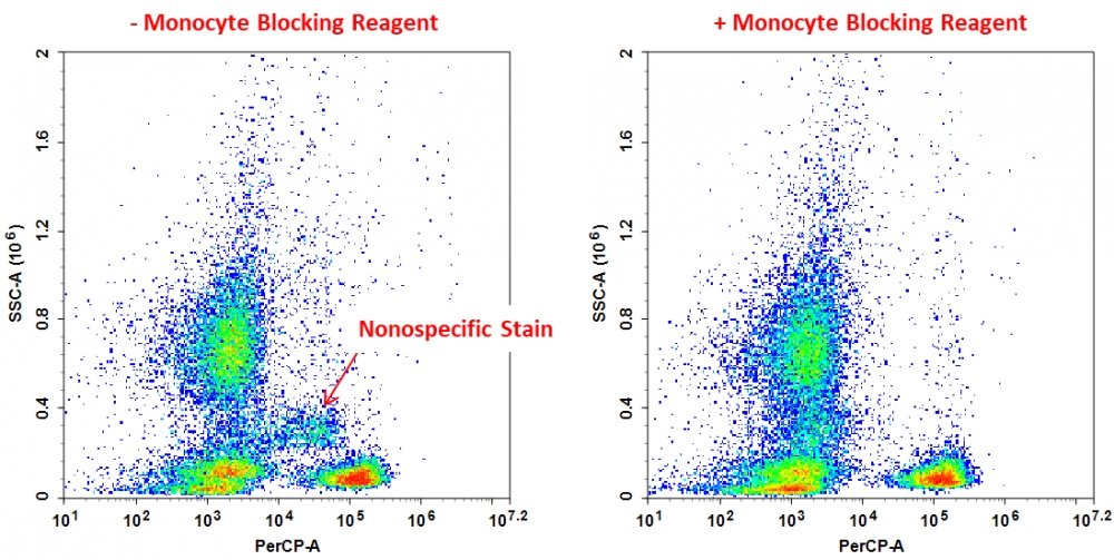
用于活细胞细胞表面染色的一些染料标记的荧光抗体缀合物通常在单核细胞和巨噬细胞中表现出非特异性结合。CytoWatch QZ100单核细胞封闭剂是一种非基于抗体的封闭溶液,经过优化可封闭基于花青的染料缀合物的非特异性背景。它可以有效地消除花青染料缀合物(例如PE / Cy5,PE / Cy7,APC / Cy7,PE / Dazzle 594,APC / Fire 750,PE / AlexaFluor®647, PE / AlexaFluor®750,APC / AlexaFluor®750,PE /iFluor®647,PE /iFluor®750和APC /iFluor®750)。该试剂对活淋巴细胞,单核细胞和粒细胞的特定表面染色没有影响。
实验方案
流式细胞仪分析的细胞表面染色方案
1.向100 µL PBMC或全血中加入5 µL CytoWatch QZ100单核细胞封闭试剂。
2.在室温下孵育5-10分钟或立即添加一抗,然后在室温下孵育20分钟。
3.用细胞染色缓冲液洗涤两次。
4.将细胞重悬于0.5 mL细胞染色缓冲液中。
5.进行流式细胞仪分析。
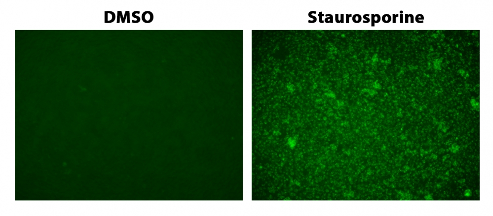
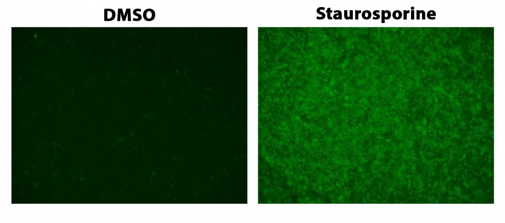
图示

图1.未经处理(左)或用CytoWatch QZ100单核细胞封闭剂(右)处理人外周血,并用CD3(克隆UCHT1)PE /Cy5染色。
参考文献
Synergistic reduction in albuminuria in type 2 diabetic mice by esaxerenone (CS-3150), a novel nonsteroidal selective mineralocorticoid receptor blocker, combined with an angiotensin II receptor blocker.
Authors: Arai, Kiyoshi and Morikawa, Yuka and Ubukata, Naoko and Sugimoto, Kotaro
Journal: Hypertension research : official journal of the Japanese Society of Hypertension (2020): 1204-1213
Neprilysin Inhibitor-Angiotensin II Receptor Blocker Combination Therapy (Sacubitril/valsartan) Suppresses Atherosclerotic Plaque Formation and Inhibits Inflammation in Apolipoprotein E- Deficient Mice.
Authors: Zhang, Hui and Liu, Gangqiong and Zhou, Wenping and Zhang, Wenjing and Wang, Kai and Zhang, Jinying
Journal: Scientific reports (2019): 6509
Prior β-blocker treatment decreases leukocyte responsiveness to injury.
Authors: Grisanti, Laurel A and de Lucia, Claudio and Thomas, Toby P and Stark, Aron and Strony, John T and Myers, Valerie D and Beretta, Remus and Yu, Daohai and Sardu, Celestino and Marfella, Raffaele and Gao, Erhe and Houser, Steven R and Koch, Walter J and Hamad, Eman A and Tilley, Douglas G
Journal: JCI insight (2019)
The Angiotensin Receptor Blocker Losartan Suppresses Growth of Pulmonary Metastases via AT1R-Independent Inhibition of CCR2 Signaling and Monocyte Recruitment.
Authors: Regan, Daniel P and Coy, Jonathan W and Chahal, Kirti Kandhwal and Chow, Lyndah and Kurihara, Jade N and Guth, Amanda M and Kufareva, Irina and Dow, Steven W
Journal: Journal of immunology (Baltimore, Md. : 1950) (2019): 3087-3102
Angiotensin II type 1 receptor blocker telmisartan induces apoptosis and autophagy in adult T-cell leukemia cells.
Authors: Kozako, Tomohiro and Soeda, Shuhei and Yoshimitsu, Makoto and Arima, Naomichi and Kuroki, Ayako and Hirata, Shinya and Tanaka, Hiroaki and Imakyure, Osamu and Tone, Nanako and Honda, Shin-Ichiro and Soeda, Shinji
Journal: FEBS open bio (2016): 442-60
Renoprotective effects of angiotensin receptor blocker and stem cells in acute kidney injury: Involvement of inflammatory and apoptotic markers.
Authors: Sherif, Iman O and Al-Mutabagani, Laila A and Alnakhli, Anwar M and Sobh, Mohamed A and Mohammed, Hoda E
Journal: Experimental biology and medicine (Maywood, N.J.) (2015): 1572-9
The TLR-2/TLR-6 agonist macrophage-activating lipopeptide-2 augments human NK cell cytotoxicity when PGE2 production by monocytes is inhibited by a COX-2 blocker.
Authors: Müller, Christina and Tufa, Dejene M and Chatterjee, Debanjana and Mühlradt, Peter F and Schmidt, Reinhold E and Jacobs, Roland
Journal: Cancer immunology, immunotherapy : CII (2015): 1175-84
Angiotensin II receptor blocker ameliorates stress-induced adipose tissue inflammation and insulin resistance.
Authors: Hayashi, Motoharu and Takeshita, Kyosuke and Uchida, Yasuhiro and Yamamoto, Koji and Kikuchi, Ryosuke and Nakayama, Takayuki and Nomura, Emiko and Cheng, Xian Wu and Matsushita, Tadashi and Nakamura, Shigeo and Murohara, Toyoaki
Journal: PloS one (2014): e116163
Azilsartan, an angiotensin II type 1 receptor blocker, restores endothelial function by reducing vascular inflammation and by increasing the phosphorylation ratio Ser(1177)/Thr(497) of endothelial nitric oxide synthase in diabetic mice.
Authors: Matsumoto, Sachiko and Shimabukuro, Michio and Fukuda, Daiju and Soeki, Takeshi and Yamakawa, Ken and Masuzaki, Hiroaki and Sata, Masataka
Journal: Cardiovascular diabetology (2014): 30
Macrophage CD14 expression in human carotid plaques is associated with complicated lesions, correlates with thrombosis, and is reduced by angiotensin receptor blocker treatment.
Authors: Hermansson, Cecilia and Lundqvist, Annika and Magnusson, Lisa U and Ullström, Christina and Bergström, Göran and Hultén, Lillemor Mattsson
Journal: International immunopharmacology (2014): 318-23
说明书
CytoWatch QZ100单核细胞封闭剂.pdf